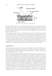
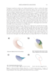
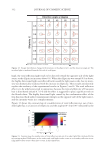
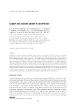
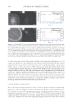
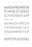
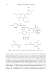
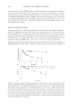
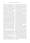
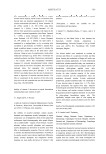
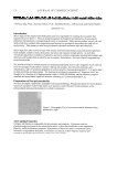
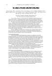
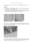
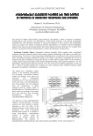
JOURNAL OF COSMETIC SCIENCE 342 Figure 2. High-resolution NanoSIMS maps from the treated Caucasian hair obtained using the Cs+ primary ion beam for 12 C2−, 32 S−, 12 C14N−, and 40 Ca16O− ions and color overlays to illustrate more clearly which signals are co-localized. The color yellow indicates the overlap of red and green, where the 32 S− and 40 Ca16O− signals are co-localized. Bar = 1 μm.

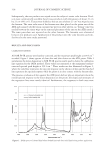
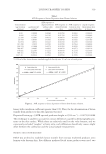
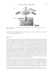
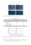
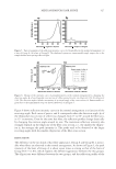
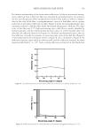
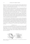
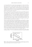
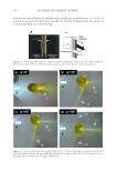
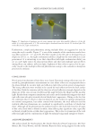
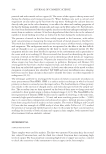
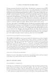
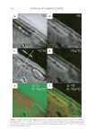
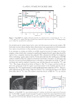
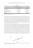
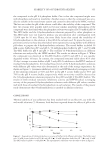
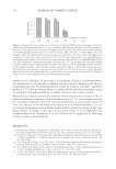
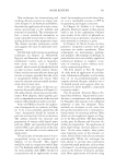
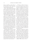
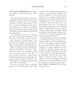
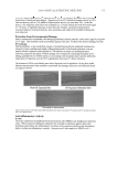
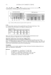
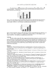
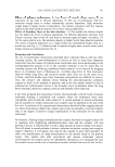
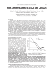
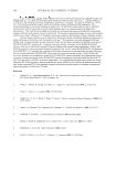
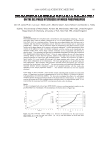
Cu AND Ca UPTAKE IN COLORED HAIR 343 the medulla and in isolated spots in the cortex in both untreated and treated samples. We had some concerns about whether these observations were representative of the extremely low copper concentration in these samples or artefacts of sample preparation. In an attempt to confi rm our ability to reliably map the location of copper ions in these samples, a hair swatch that contained a signifi cantly higher level of copper than would be expected in a consumer situation was prepared. The colorant-treated hair was washed in water containing 1 ppm copper ions for 30 min until the hair was visibly green. ICP-MS analysis confi rmed that the copper level had increased from 120 ppm to 11,000 ppm and that the calcium level had dropped from 8,700 ppm to 4,800 ppm (see Table I). The O− ion beam was used for analysis of positive ions, and the detector was tuned to 40 Ca+ and 63 Cu+. The images obtained are shown in Figure 4. The 40 Ca+ image is similar to what we have seen previously, with the signal concentrated both in the medulla and the cuticle. However, the 63 Cu+signal intensity is still signifi cantly lower than that of 40 Ca+. One pos- sible explanation for this result is that the yield of 63 Cu+ ions from the hair is very much lower than that of 40 Ca+ ions. The relative sensitivity factor, or RSF, for 40 Ca+ in a silicon Figure 3. NanoSIMS line profi les obtained using the Cs+ primary beam on treated hair for 12 C2−, 32 S−, 12 C14N−, and 40 Ca16O− signals. The line scan direction is indicated with an arrow in the 12 C14N image in Fig- ure 2a. Abbr: Ex, exocuticle En, endocuticle. Figure 4. (a) NanoSIMS 40 Ca+ map obtained using the O− primary ion beam from copper-treated Caucasian hair. (b) NanoSIMS 63 Cu+ maps obtained using the O− primary ion beam from copper-treated Caucasian hair. Bar = 10 μm. (c) NanoSIMS line profi les of the 40 Ca+ and 63 Cu+ signal obtained using the O− primary ion beam on copper-treated Caucasian hair. The line scan direction is indicated with an arrow in the 40 Ca+ image in (a).
Purchased for the exclusive use of nofirst nolast (unknown)
From: SCC Media Library & Resource Center (library.scconline.org)