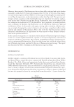
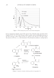
EVALUATION OF ANTI-CELLULITE EFFICACY 307 the capillary wall and preventing fl uid leakage (12). The pharmacological activity is then a decrease in capillary permeability, and an anti-edema effi cacy is observed. Escin also possesses signifi cant anti-infl ammatory properties, which can, however, be hardly separated from the anti-edema effi cacy on pharmacological models. The combination of the mechanisms of action identifi ed so far appoints escin as a relevant active ingredient in the treatment of cellulite. MATERIALS AND METHODS FORMULATION The formulation of the topical product (ACTIVE) was developed with Sinerga Research Center Laboratories (Pero, Italy). The development took into account not only the effi cacy of the active substances to counteract the causes of cellulite blemishes, but also the pleas- antness of the fi nal formulation in order to achieve the best possible compliance. The botanical active ingredients formulated in ACTIVE are visnadine (0.25% w/w), Ginkgo biloba Dimeric Flavonoids Phytosome®(0.5% w/w), and escin (1% w/w). The association of these active ingredients (described in patent WO2005/004858) has been selected accord- ing to the specifi c profi le of each single substance. TRIAL DESIGN The aim of the study was to clinically assess the cosmetic effi cacy of a topical treatment to be applied on the thighs over a period of four weeks. Morphometric analysis and in- strumental evaluations were carried out. Twenty-fi ve female volunteers (ages: 30–55 yrs), affected by fat accumulations and/or slight-to-moderate edematous-fi brosclerotic pan- niculopathy in the lower limbs, were selected for the study. The multifunctional product ACTIVE, containing the botanical ingredients visnadine (0.25%), Ginkgo biloba Dimeric Flavonoids Phytosome® (0.5%), and escin (1%) (obtained from Indena S.p.A., Italy), was compared to its relevant placebo formulation PLACEBO. All volunteers provided a writ- ten informed consent. The trial was conducted in a single-blind method with the com- parison within subjects (each subject being its own control), and volunteers were required to apply the test products on the thigh twice a day, unilaterally, for a period of four con- secutive weeks. They underwent two medical examinations, a baseline evaluation at T0 before the beginning of the test, and an evaluation at the end of the treatment period T4. Body weight, in the case of substantial differences between the beginning and the end of the trial, was considered as a dropout criterion. However, no dropouts due to relevant body-weight variations occurred (during the course of the study, the two dropouts quit the trial due to personal reasons independent from the trial itself). Additionally, at the beginning of the treatment, the overall tolerability of the treatment was also observed. Finally, the effi cacy of the treatment was assessed both clinically and instrumentally. CLINICAL EVALUATION The clinical assessment was carried out at the upper, median and lower third of the thigh, according to the following evaluations:

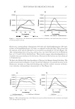
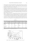
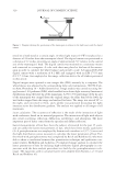
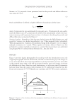
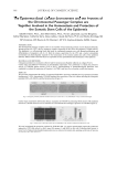
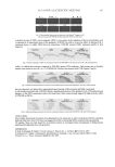
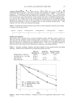
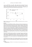
JOURNAL OF COSMETIC SCIENCE 308 (a) Visual clinical score for assessment of the degree of cellulite (both visual appearance and appear- ance at pinching on the basis of a specifi c photographic reference scale): 0–no observable cellulite 1–no panniculus, some mild depressions 2–some panniculus separated from mild de- pressions 3–several panniculus, separated from medium depressions and 4–broad pan- niculus separated from deep depressions (0 and 4 were considered exclusion criteria). (b) Firmness of the inner thigh (0–very low 1–low 2–moderate 3–good 4–very good). (c) Skin smoothness (0–very low 1–low 2–moderate 3–good 4–very good). (d) Degree of pain at pinching (0–absent 1–mild 2–moderate 3–severe 4–very severe). INSTRUMENTAL EVALUATION The instrumental evaluations were carried out through: (a) Contact thermography for the assessment of the thermographic stage of cellulite (13). At each control, contact thermography was conducted by means of liquid crystal thermographic plates that can detect a temperature range form 28° to 34°C through color visualization (from the coldest to the warmest: black–brown–yellow–green–light blue–pink–dark green–blue). The clinical meaning of contact thermography refl ects the amount of heat that is transmitted to the plate by the contact with the skin. The skin temperature is a clear indication of cutaneous microcirculation, and its variations allow assessing the effi - cacy of vasokinetic treatments: skin temperature increases in the case of vasodilation, in- creased number of open capillaries, and increased local metabolism, while it decreases following vasoconstriction, a decreased number of blood vessels, and a fat tissue increase. One thermographic aspect typical of panniculopathy is dysthermia, with wide hypother- mic areas. The panniculopathy evaluation was based upon the following classifi cation: 0–homogeneous “warm” aspect of the thermographic image 1–dysthermiae 2–“venous” lakes 3–wide cold areas and 4–“cold” aspect of the thermographic image. (b) Morphometric measurements of thigh circumferences (upper, median, and lower third). All cir- cumferences were measured in standardized conditions at the upper, median, and lower third levels of the thigh thanks to a specifi c electro-optical system able to defi ne the vol- unteer’s position. Measurements were taken three times for each site both single values and the median value were recorded. The electro-optical system (composed of a support, a horizontal bar with two laser beams, and a graduated panel in front of the support) al- lowed us to establish precisely the volunteer’s position with respect to the graduated panel behind him (1 mm approximation). In order to determine the coordinates, the subject stood in front of the graduated panel and the operator drew the feet position in order to put him back in the same position during the following visits. Then the laser beam was set to tangentially touch the volunteer’s leg and was pointed on the graduated panel. The same point was then marked on the volunteer’s skin (by means of a dermo- graphic pencil) and the measurement of the thigh circumference was taken. (c) Skin plastoelasticity measured on the inner thigh for the evaluation of elasticizing/fi rming effi - cacy (14). The evaluation of skin plastoelasticity was carried out at the inner thigh level by the means of a dermal torque meter (Diastron LTD). The instrument applies the tech- nique of in vivo torsion by a probe composed of two concentric circles 3 mm apart. The inner circle, spinning slowly, determines a constant torsion on the skin. When the skin resistance reaches 9mNm, the torsion stops. The torsion time is 1 second. The equipment
Purchased for the exclusive use of nofirst nolast (unknown)
From: SCC Media Library & Resource Center (library.scconline.org)