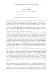
JOURNAL OF COSMETIC SCIENCE 36 Green tea contains polyphenolic compounds, mainly catechin, which is further composed of several monomers including epicatechin, epicatechin gallate, epigallocatechin (EGC), and epigallocatechin-3-gallate (EGCG). Among all the monomers, EGCG has the highest proportion, accounting for about 50% of catechins (4,5). It has a strong antioxidant activ- ity and can effectively remove the active free radicals within human body, inhibit lipid peroxidation (LPO), and reduce oxygen free radical (OFR) and LPO-induced damage to cells, DNA, or other biological macromolecules (6–8). Through its strong antioxidant effects, EGCG can also reduce the intracellular reactive oxygen species activity, block the ultraviolet (UV) radiation, inhibit the matrix metalloproteinase activation and collagen damage on human dermal fi broblasts, and increase the extracellular matrix (9–11). Exogenous sources for the production of cellular hydrogen peroxide (H2O2), especially in the skin, are UVA and UVB irradiation (12). In the study of skin photoaging, the most common skin lesions are caused by chronic UV irradiation, which involves the demise, aging, and apoptosis of human dermal fi broblasts. Therefore, the establishment of an in vitro model of H2O2-induced human dermal fi broblasts apoptosis is crucial for future studies on the oxidative damage of human skin, screening of antioxidants, and its application in the cosmetics. Although many studies have reported that EGCG has strong antioxidant activity (13,14), little is known about its role in the model of H2O2-induced human der- mal fi broblast apoptosis (15). In this study, for the fi rst time, we address the effect of EGCG on oxidative damage and apoptosis induced by H2O2 in human dermal fi broblasts and explore the mechanism of its protective role, which provides a theoretical basis for the application of EGCG in cosmetics. In this study, we used H2O2-induced oxidative damage in human dermal fi broblasts as a model to study the effects of EGCG on H2O2-induced oxidative damage and apoptosis and examined its underlying mechanism. MATERIALS AND METHODS MATERIALS Dulbecco’s modifi ed Eagle’s medium (DMEM) and fetal calf serum (FCS) were purchased from Gibco Life Technologies (Grand Island, NY). H2O2 (3%), 2,2-diphenyl-1-picrylhydrazyl (DPPH), Propidium Iodide (PI), and EGCG (purity 98%) were purchased from Sigma-Aldrich (St. Louis, MO). Terminal deoxynucleo tidyl tran sferase dUTP nick end labeling (TUNEL) apoptosis detection kit was purchased from Roche (Basel, Swit- zerland). Superoxide dismutase (SOD), malondialdehyde (MDA), and glutathione per- oxidase (GSH-px) detection kit were purchased from Jiancheng Biological Engineering Academy (Nanjing, China). CELL CULTURE Primary cultures of skin fi broblasts from a healthy boy’s foreskin left over from surgery were grown on plastic fl as ks under standard conditions: DMEM supplemente d with 10% CS at 37°C in a humidifi ed atmosphere of 5% CO2. According to the different pur- poses of experiments, cells were quantitatively planted onto the cell culture bottles or 30-mm petri dishes for future experiments. Third- to sixth-generation cells were used for

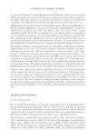
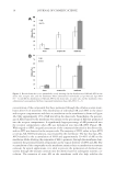
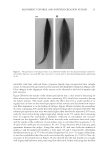
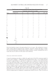
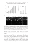
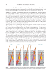
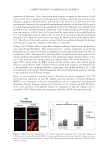
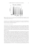
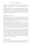
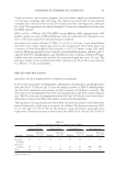
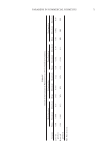
EFFECT AND MECHANISM OF EGCG AGAINST H2O2-INDUCED OXIDATIVE DAMAGE IN HUMAN DERMAL FIBROBLASTS 37 experiments. For each experiment, we used a seeding density of 5 × 10⁴ cells/well plated onto 30-mm petri dishes. The cells were grown to 70% confl uency over 24 h. MTT ASSAY Cell suspensions were made in fi bro blast growth medium containing different drug con- centrations with a density of 5 × 10⁴ cells/ml. Cells were plated at a density of 5 × 10³ cells/well in 96-well plates (200 μl/well, equivalent to 1 × 10⁴ cells/well). Each different concentration of drugs occupied six holes, repeated twice, and incubated at 37°C with 5% CO2. 3-(4,5-dimethylthiazol-2-yl)-2,5-diphenyltetrazolium bromide (MTT) (20 μl/ well) was added to each hole 4 h before the experiment ended. The supernatant was dis- carded 4 h later. Finally, 150 μl dimethyl sulfoxide was added and shaken for another 15 min until the crystals were completely dissolved. The absorbance was measured at a wavelength of 570 nm (A570) by a microplate reader. Figure 1. Oxidation damage to human dermal fi broblasts induced by different concentrations of H2O2. (A) Statistical analysis of cell death rate measured by an MTT colorimetric assay with different concentrations of H2O2 and treatment times. *p 0.05 according to one-way ANOVA. Data are obtained from fi ve indepen- dent experiments. (B) Cell viability and damaged cell nuclei detected by Hoechst staining and TUNEL assay after exposure of human dermal fi broblasts to different concentrations of H2O2. The scale bar represents 100 μm. (C) Statistical analysis of TUNEL-positive cells resulting from treatments with different concentrations of H2O2. *p 0.05 according to one-way ANOVA. Data are obtained from fi ve independent experiments.
Purchased for the exclusive use of nofirst nolast (unknown)
From: SCC Media Library & Resource Center (library.scconline.org)