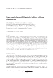
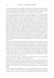
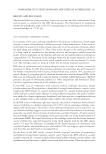
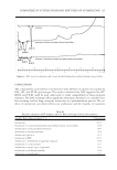
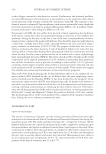
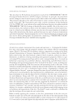
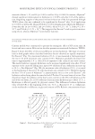
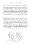
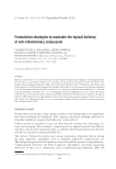
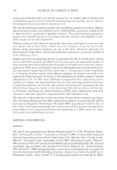
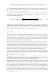
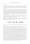
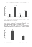
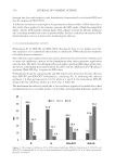
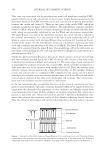
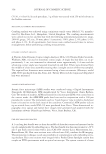
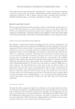
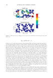
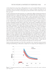
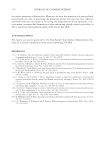
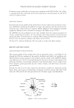
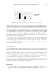
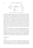
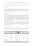
JOURNAL OF COSMETIC SCIENCE 356 They carry out a structural role by providing hair with a cell membrane complex (CMC), present in both cuticle and cortical cells. In recent years, insight has been gained as to the structural details of the CMC for both cuticle and cortical cells as well as the interface between the cuticle and cortex (1). There are two parts of the cuticle CMC, which are normally classifi ed as the upper and lower β layers. The upper β layer contains covalently- bound 18-MEA, which is believed to form a monolayer interspersed with other free fatty acids, which are presumably stabilized by van der Waals and electrostatic interactions. The upper β layer is located on the uppermost lamina of the cuticle cell and is exposed to the external environment. It is also present at the top of each underlying cuticle cell where it comes in contact with the lower β layer of an overlying cell. Located on the un- derside of the cuticle cell, the lower β layer consists of a monolayer containing free fatty acids and covalently attached fatty acids (but, no 18-MEA). The lower β layer of an over- lying cell is separated from the upper β layer of an underlying cell by the delta layer, in- tercellular cement holding the two cells together, which is thought to be glycoprotein or globular protein (Fig. 1). Unlike the upper and lower β layers in the cuticle, which contain a mixture of covalently and noncovalently attached lipids, the CMC of cortical cells consists of free fatty acids, cholesterol (or cholesterol sulfate), and ceramide (1). The outer edge of each cortical cell is surrounded by a bilayer structure, the cortical CMC, which is further enveloped by a delta layer that acts as the interface with another cortical cell. Thus, two bilayers from adjacent cortical cells are separated by a thin delta layer (Fig. 2). The interface between cuticle and cortical cells is a composite CMC composed on the cuticle side of a mixed monolayer of covalently and noncovalently attached fatty acids (lower β layer) and on the cortical side of a bilayer of free fatty acids, cholesterol, and ceramide. The cuticle mono- layer and cortical bilayer are also separated by a delta layer. In the last several decades, considerable attention in the research community has been given to structural lipids, especially covalently bound 18-MEA (2–4). Lipids of sebaceous origin were often deemed as less important, or even a nuisance, and thought to play little or no functional role in hair. In skin, sebum is an integral component that carries out a protective functional role. Sebaceous secretions provide a lipid-rich hydrophobic sub- stance that protects the outermost surface of skin, thereby enhancing barrier function. They also transport antioxidants and antimicrobial peptides to the skin surface to protect Figure 1. Schematic of the CMC (lower and upper β layers) of two overlying cuticle cells. The lower β layer corresponds to the overlying cell whereas the upper β layer corresponds to the underlying cell. Reprinted with permission from Society of Cosmetic Chemists. Copyright 2009. Originally appeared in Reference 1.

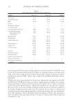
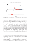
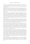
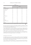
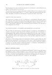
PHYSICOCHEMICAL PROPERTIES OF DELIPIDIZED HAIR 357 it from environmental insult (e.g., UV radiation) and invasion by foreign pathogens. Se- baceous lipids consist of free fatty acids, triglycerides, free cholesterol, cholesterol and wax esters, paraffi ns, and squalene. In this work, we investigated the physicochemical properties of delipidized hair using cutting-edge instrumental techniques to probe the surface and interior of the fi ber struc- ture. During our extraction procedure, we removed both sebaceous lipids and free struc- tural lipids from the exterior and interior of the fi ber. For this reason, it is diffi cult to discern differences in the structural/functional contributions of measured fi ber properties by either sebum or free structural lipids. It should be noted though that we do not re- move covalently attached lipids (e.g., 18-MEA) from hair. Therefore, any changes we observe in the physicochemical properties of delipidized hair by solvent extraction are due to free structural lipids or lipids of sebaceous origin. MATERIALS AND METHODS Studies were conducted on virgin and delipidized hair. We removed noncovalently at- tached lipids from hair using a Soxhlet extraction technique. Various instrumental tech- niques were used to examine differences between virgin and delipidized hair and to discover the importance of these lipids in the physicochemical behavior of hair. Details of each experimental technique are provided in the following sections. DELIPIDIZATION OF HAIR Using a Soxhlet extraction apparatus, free internal and surface lipids were removed from hair. This method is based on an established procedure in which hair is extracted with a series of solvents of increasing polarity (5). The apparatus consisted of a round bottom fl ask to which a Soxhlet extraction tube was mounted. Inside the Soxhlet extraction tube, a bundle of hair was placed in a cellulose thimble. A condenser was mounted on top of the Soxhlet extraction tube. The effect of solvent extraction on hair was investigated fi rst by treat- ment with t-butanol and n-hexane for 4 h each, then with a mixture of chloroform/methanol Figure 2. Schematic of the cortical CMC of two adjacent cortical cells separated by a delta layer. Reprinted with permission from Society of Cosmetic Chemists. Copyright 2009. Originally appeared in Reference 1.
Purchased for the exclusive use of nofirst nolast (unknown)
From: SCC Media Library & Resource Center (library.scconline.org)