J. Cosmet. Sci., 66, 285–293 (September/October 2015) 285 The top echorich band in a 50-MHZ ultrasound sonogram refl ects epidermal properties DI QU and SARAH WHITEHEAD, Research & Development, Amway Corporation, Ada, MI 49355. Accepted for publication July 7, 2015. Synopsis High-frequency ultrasonography is a useful noninvasive tool to measure the acoustic properties of skin. Due to the ambiguity or confusion over the meaning of the skin entry echo, measurements have been limited to the dermis or full skin thickness with little data on epidermal properties. The purpose of this study was to better understand the nature of the skin entry echo and determine whether it is related to epidermal struc- ture. We approached the problem by dampening the sudden change in material density from the coupling medium to the skin surface using facial tissue as a masking material. The thickness and acoustic density of bare and masked skin sites were measured using dermal ultrasound with a 50-MHz transducer. Results showed that the original thickness and acoustic density of the skin entry echo did not change when the skin was masked up to two layers. A comparison between the epidermal thicknesses measured using ultrasound and confocal microscopy also indicated that the two methods yielded about the same results with no statisti- cally signifi cant difference detected. This study demonstrates that the purported skin entry echo is not just a meaningless artifact, and it refl ects useful properties of epidermal structure. INTRODUCTION Since fi rst reported in 1979 (1), dermal high-frequency ultrasonography has been shown to be a useful noninvasive tool to understand skin properties. Numerous studies have focused on measuring skin thickness at various anatomical sites from people of different ages, genders, ethnicities, and disease states (2–7). Due to ambiguity or confusion over the meaning of the skin entry echo, many of these measurements were limited to the dermis only or to the full skin thickness, with very little data on the thickness and the echogenicity of the epidermis. It is evident in the literature that such ambiguity or confusion exists. Most researchers be- lieve that the skin entry echo is an artifact caused by the sudden change in impedance be- tween the coupling medium and the stratum corneum, which makes the epidermis diffi cult to visualize or measure using ultrasonography (8,9). El Gammal et al., using a 100-MHz The study was originally presented at the U.S. Symposium of ISBS (2002), Baltimore, MD. Address all correspondence to Di Qu at Di.Qu@amway.com.

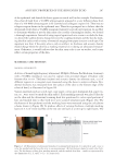
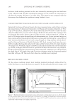
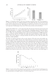
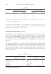
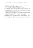
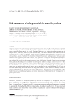
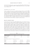
JOURNAL OF COSMETIC SCIENCE 286 transducer to visualize normal and psoriatic skin, suggested that for normal glabrous skin, the stratum corneum was sonographically invisible and the fi rst echorich band seen in the sonogram was an artifact and deemed as the entry echo. The viable epidermis and papillary dermis were represented by an echopoor band beneath the entry echo. They further stated that the entry echo remains, no matter whether the horny layer is stripped, occluded with topical agents, or entirely removed (10). Other researchers consider the entry echo band to represent the epidermis without providing adequate supporting data, and have reported correlations between its echogenicity and epidermal hydration or infl ammatory states (11–15). Nouveau-Richard et al. (16) compared their measurement results between confocal laser scanning microscopy and the dermal ultrasound with a 20-MHz transducer, and con- cluded that the distance between the fi rst two hyperechoes on an ultrasound A-Scan sono- gram represented the epidermal thickness. Their study had provided us with confi dence to conduct this study to further support the argument that the sonograms of dermal ultra- sound, at least those obtained by using a 50-MHz transducer, contain information to refl ect the properties of the epidermis of human skin. In regard to the epidermal thickness measurement, skin histology has been considered as the “gold standard”. Sandby-Moller et al. (17) conducted an extensive study to examine the epidermal thickness from 71 Caucasians, and reported that the total epidermal thickness (stratum corneum plus the cellular epidermis) to be about 83.7 (±16.6) μm. This result serves as a good reference to validate the measurement results of instrumental, in vivo, non- invasive methodologies although tissue shrinkage in biopsy samples was a concern (10). In a typical 50-MHz ultrasonogram of normal human glabrous skin, there exists a clear, well- defi ned echorich band on the top section of the B-Scan image. Its thickness is approximately 100 μm, the typical thickness of the epidermis, suggesting there is a possibility that the echorich band may refl ect the epidermal thickness. Figure 1A shows a comparison between the images of the histology of human normal skin (18) and the ultrasound B-Scan sono- gram. It demonstrates the geometric similarity between these two methods, and the scales Figure 1. (A) Comparison of skin thickness scales between images of histology and the dermal high-frequency ultrasound. In the histological image, L1 = epidermis and L2 = dermis. In the ultrasound image it shows a B-scan sonogram of normal human skin. [Source of histology: School of Anatomy and Human Biology, The University of Western Australia (18).] (B) Graphical representation of epidermal thickness measurement method in DUBplus software. The location of the red vertical line is generally set to include the bottom of the echorich band.
Purchased for the exclusive use of nofirst nolast (unknown)
From: SCC Media Library & Resource Center (library.scconline.org)