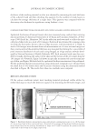
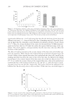
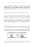
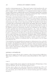
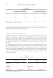
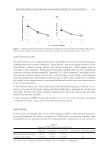
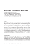
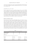
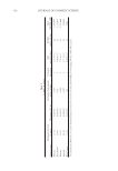
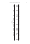
ACOUSTIC PROPERTIES OF THE SKIN ENTRY ECHO 291 Figure 7. Distributions of the measured epidermal thickness of human skin. (A) Distribution of epidermal thickness of cheek skin by using ultrasound with a 50-MHz transducer. N = 102, mean = 104 μm, and stan- dard deviation (SD) =11.9 μm. (B) Distribution of epidermal thickness of dorsal and ventral forearm skin using confocal laser scanning microscope. N = 25, mean = 108.2, and SD=16.8 μm. corresponding change in acoustic density after the skin was masked since the sudden shift in impedance from the coupling medium to the surface of skin had been dampened. In other words, the acoustic density of the original skin entry echo would have been dramatically reduced if the impedance change from the masking material to the skin surface had become much less. Therefore, this result further supports our argument that the skin entry echo, commonly purported to be an artifact, may represent properties of the epidermis. COMPARISON BETWEEN ULTRASONOGRAPHY AND CONFOCAL LASER SCANNING MICROSCOPY The epidermal thickness measured using confocal microscopy was very similar to that of the ultrasound method. The average thickness value obtained from 10 people (25 skin sites of dorsal and ventral forearms) was 108.2 (±16.8) μm while the value measured from the sonograms of ultrasound was 104.6 (±11.9) μm. Plotting the results of epidermal thickness measured using ultrasound method from the 102 volunteers in a histogram we obtained a quite normal distribution. The thickness values ranged from 70 to 150 μm with the mode of the distribution falling between 100 and 110 μm as shown in Figure 7A. Comparing this chart to the results obtained using confocal microscope in Figure 7B we can see that these two distributions are very similar. Student’s t test on these two data sets showed no statistical difference between them (p = 0.329), and therefore it is diffi cult to believe that it is just a coincidence when these two sets of data match so well. DISCUSSIONS ABOUT THE TECHNIQUE The approach illustrated in this study helped us to collect experimental evidence and to show that the skin entry echo may refl ect the structural information of skin, not just a meaningless artifact. Use of paper tissue to mask the surface of skin was found to be a convenient way to test the argument that the thickness and pixel intensity should have been changed had the skin entry echo been an artifact caused by the sudden change in echoing property from coupling water to the skin. While the physical masking method

JOURNAL OF COSMETIC SCIENCE 292 works, other approaches would be more direct to validate ultrasound measurement of epidermal thickness. One of the approaches would be an ex vivo method which measures the thickness directly from an epidermal sheet separated from the cadaver skin using enzyme digestion methodology. We will examine the feasibility of this approach as the next step to this investigation. Additionally, the relevance of this technique to the 20-MHz transducer would need to be addressed. Evolved in the past decades, 20 MHz has been a standard method of ultrasound measurement of skin. When we designed this study, we had chosen the 50-MHz transducer since it had better sonogram pixel resolu- tion and the imaging depth was well within the full thickness of human skin. The more detailed view of rete ridge structure of epidermis in 50 MHz provided us with the ability to more accurately detect the minimum thickness of epidermis (from skin surface to the top of the rete ridge structure) particularly when it is required to compare the results obtained from the confocal microscope. A 20-MHz transducer had resulted in sonograms with a more uniform boundary at the edge of the skin entry echo, which had yielded epidermal thicknesses toward the higher end when compared to the biopsy and confocal results. Adjusting the gain level may be able to improve the resolution and it needs a thorough investigation as the next step to establishing proper conditions which work with transducers of various frequencies. CONCLUSION The results of this study demonstrate that the acoustic properties of the skin entry echo correlates to epidermal thickness. Our observations indicate that the presence of the echorich band beneath one or two masking layers is consistent in thickness with the echorich band of adjacent bare skin. The acoustic density and thickness of the purported skin entry echo remained the same despite being masked. If the echorich band known as the skin entry echo is an artifact due solely to a sudden impedance change, the thickness and acoustic density would change accordingly once the skin is masked. In this experi- ment no drastic changes in acoustic density or thickness were observed after the skin was masked. Thus, researchers should not avoid using this echorich band in 50-Mhz ultra- sound sonograms to study relative changes in the thickness or acoustic density of the epidermis. Understanding that the ultrasound method may not have the resolution as high as confocal microscope for accurate measurement of epidermal thickness, this result may provide us with a rough estimate of epidermal properties when other more advanced techniques or methods are not available. REFERENCES (1) H. Alexander and D. L. Miller, Determining skin thickness with pulsed ultrasound, J. Invest. Dermatol., 72, 17–19 (1979). (2) J. Waller and H. Maibach, Age and skin structure and function, a quantitative approach (I): blood fl ow, pH, thickness and ultrasound echogenicity, Skin Res. Technol., 11, 221–235 (2005). (3) M. Gniadecka, Effects of ageing on dermal echogenicity, Skin Res. Technol., 7, 204–207 (2001). (4) C. Lasagni and S. Seidenari, Echographic assessment of age-dependent variations of skin thickness: a study on 162 subjects, Skin Res. Technol., 1, 81–85 (1995). (5) M. Gniadecka, J. Serup, and J. Sondergaard, Age-related diurnal changes of dermal oedema: evaluation by high-frequency ultrasound. Br. J. Dermatol., 131, 849–855 (1994).
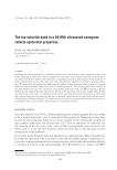
Purchased for the exclusive use of nofirst nolast (unknown)
From: SCC Media Library & Resource Center (library.scconline.org)