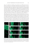
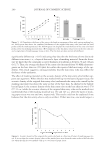
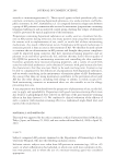
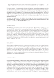
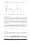
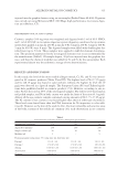
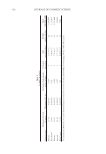
ACOUSTIC PROPERTIES OF THE SKIN ENTRY ECHO 289 they were considered not applicable to the study. Paper tissue (Kleenex® facial tissue), however, was found to be a meaningful masking agent, which we believed to have re- fl ected the sound waves the similar way as that of the cellular epidermis due to its porous structure when soaked in the coupling water during a measurement. The ultrasonogram of skin with a single mask layer placed on it is shown in Figure 4A in which the thickness of the entry echo, T, is nearly doubled at the masked site on the right-hand side of the image when compared to that of the bare skin, X, on the left-hand side. We can see that this increased thickness is a combination of the original entry echo of the skin and that of the masking material on top of it. Figures 4B–I show the sonograms, especially the changes in the entry echo thickness of the masked skin sites. With additional layers of masking material placed on skin there was a corresponding increase in thickness T. The thickness of the entrance echo from the masked and unmasked skin sites was mea- sured from Figures 4B–I. For the bare skin site, it had an average value of 116.0 ± 9.5 μm. To determine the thickness of each masking layer, we fi rst subtracted X from T in each of the eight sonograms to obtain a total thickness of the masking layers on skin. Cumula- tive thickness curves when plotted against the number of masking layers are shown in Figure 5A. Then, dividing this total thickness by the number of masking layers in each sonogram, the thickness of a single masking layer was calculated. From the eight sonograms shown in Figure 4, the average thickness of a single masking layer was 90.7 ± 4.5 μm. Figure 5B shows the thickness values of the entry echo measured from the bare skin sites as well as from the masked sites. These two populations were found to be Figure 4. (A) Ultrasound sonogram of ventral forearm skin masked with a layer of paper tissue on the right- hand side showing a wider entry echo band than that on the left-hand side of the image. (B–I) Sonograms after rotating 90° counterclockwise and a phasing operation to align the surface to a vertical line. The thick- ness of entry echo band increased with increasing layers of masking tissue, from one layer (B) to eight layers (I). The upper half of each image shows the masked skin and the lower half is bare skin. T = total thickness of entry echo X = bare skin entry echo.

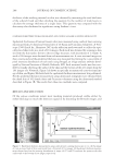
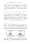
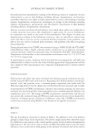
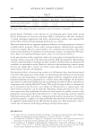
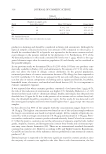
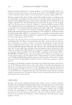
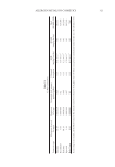
JOURNAL OF COSMETIC SCIENCE 290 signifi cantly different (p 0.05) indicating they describe the thickness of two distinctly different structures, i.e., a layer of skin and a layer of masking material. From the litera- ture we know that the commonly accepted thickness of epidermis is between 70 and 140 μm (16,17). Thus, the average thickness of the entry echo measured from 50-MHz ultrasono- grams on the bare skin site (116 μm) fi ts within the reported thickness range of the epi- dermis. This result suggests a strong possibility that the skin entry echo may refl ect the thickness of the epidermis. The effect of masking material on the acoustic density of the skin entry echo further sup- ports our argument. When the skin was masked with up to two layers of paper tissue, the acoustic density of the original skin entry echo (underneath the entry echo caused by the masking material) was about the same as that of the bare skin. As shown by the hollow bar in Figure 6, the acoustic density of the skin entry echo on the bare skin site was 147.8 (±7.13 a.u.) while the acoustic density of the original skin entry echo on the masked sites (underneath that of the masking material) was 152 and 145 a.u. when the layers of mask- ing paper tissue was one and two, respectively. This result could not be explained if one believes that the skin entry echo is only an artifact. If that was true, one would expect a Figure 5. (A) Cumulative thickness of the entry echo band in sonograms of skin with various masking lay- ers. Solid square line shows the combined thicknesses of the original skin entry echo band and the band produced by the masking material. The hollow square line displays the total thickness of the entry echo band produced by the masking material alone. (B) Comparison of the thickness values between the bare skin site and a single layer of masking paper tissue on skin. The two data sets were statistically different. Figure 6. Acoustic density of the original skin entry echo band as a function of layers of masking paper tis- sue. It is seen that the acoustic density values of the masked sites were about the same as that of the bare skin site when the skin site was masked with up to two layers of paper tissue.
Purchased for the exclusive use of nofirst nolast (unknown)
From: SCC Media Library & Resource Center (library.scconline.org)