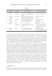
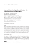
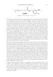
JOURNAL OF COSMETIC SCIENCE 164 (3) draw attention to the possibility of bacterial contamination of cosmetic tools and products in the case of when they are used by a person with a bacterial infection. Disi nfection, i.e., the deliberate use of chemical and physical factors to reduce the num- ber of biological pathogenic microorganisms, contributes to the elimination of vegetative forms of microorganisms. However, the spores of these microorganisms are normally resistant to chemical disinfectants. Appropriate disinfection of surfaces and tools in a beauty salon prevents the spread of infection from one client to another. The correct selection of disinfectant plays an important role in the disinfection process, as different disinfectants have different effi ciencies. There are many reports in the literature regarding disinfection in medicine and dentistry, but there are no reports on this subject in the fi eld of cosmetology (4). This research was conducted to increase knowledge of the processes of choosing effective disinfection methods in a beauty salon. The aim of the study was to compare the effective- ness of three different preparations for disinfecting reusable make-up applicators with regard to their duration of action to inhibit microbial growth. MATER IALS AND METHODS The f ollowing reusable make-up applicators were subjected to the research: the natural make-up brush M Brush by Maxineczka No. 07 (Maxineczka, Lódź, Poland), the syn- thetic make-up brush Zoeva No. 104 (ZOEVA GmbH, Frankfurt am Main, Germany), and also the Beauty Blender make-up sponge (Rea.deeming Beauty, Inc., Bethlehem, PA). Before starting the tests, all the applicators were used for applying face makeup. The e ffectiveness of the agents was assessed based on the change in the number of micro- organisms present before and after disinfection. The analysis of the number of microor- ganisms present was performed using the plate method. DISIN FECTANTS Commo nly used disinfectants were used to disinfect the applicators: 1. Pr otex Ultra bactericidal soap from Colgate-Palmolive (New York, NY), 2. 70 % medical spirit (ethanol) from Alpinus Chemia (Solec Kujawski, Poland), batch number: 1178D120718, series No.: 1179A160718, 3. Hy dro Sept solution for disinfecting surfaces and tools from Silcare Company (Gorzów Wlkp., Poland), permission to trade biocidal product No.: 2072/05, series No.: 08/2019. LABOR ATORY MATERIAL The f ollowing were used in the tests: 1. Si x sterile ready-to-use Sabouraud-Gentamicin Chloramphenicol two agar from BioMerieux Co. (Marcy l'Etoile, France) containing agar on Petri dishes with a diameter of 90 mm (Solution Basin, 55 mL, from Heathrow Scientifi c, Vernon Hills, IL) 2. On e sterile plastic pipette, 3. Th ree sterile plastic bacteriological tubules from Thermo-Scientifi c (Waltham, MA), 4. On e 250-mL glass beaker, 5. Si ngle-use gloves,

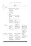
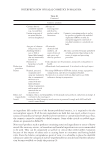
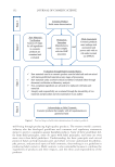
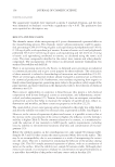
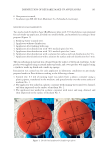
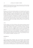
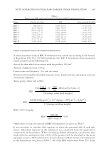
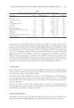
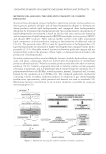
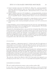
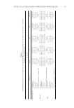
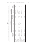
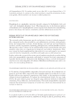
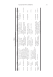
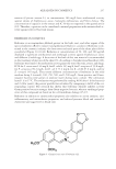
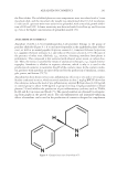
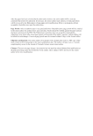
DISINFECTION OF REUSABLE MAKE-UP APPLICATORS 165 6. On e protective mask, 7. In cubator type BE 400 from Memmert Co. (Schwabach, Germany). MICRO BIOLOGICAL EXAMINATION Two r eady-made Columbia Agar (BioMerieux) plate with 5% blood plates were prepared for each make-up applicator, divided into smaller fi elds, and numbered according to their purpose (Figure 1): 1. Fr esh tap water (control test). 2. Ap plicator without disinfection. 3. Ap plicator after washing with soap. 4. Ap plicator after disinfection with 70% medical spirit for 30 s. 5. Ap plicator after disinfection with 70% medical spirit for 3 min. 6. Ap plicator after disinfection with a solution for surface and tool disinfection for 30 s. 7. Ap plicator after disinfection with a solution for surface and tool disinfection for 3 min. The m icrobiological material was obtained from the surface of the facial epidermis. A dry product was applied using a natural make-up brush, and a wet product was applied using a synthetic make-up brush and a make-up sponge. Inocu lation was carried out for each applicator in laboratory conditions on previously prepared media in Petri dishes according to the following scheme: 1. Co ntrol test: 0.5 mL of running water was taken from a plastic container using a plastic pipette, transferred to the medium, and spread evenly over the entire surface of medium No. 1. 2. Th e applicator was soaked in a plastic container with running water until wet, drained, and then impressed on the surface of medium No. 2. 3. Th e applicator was washed in a plastic container with water and soap, drained, and then impressed on the surface of medium No. 3. Figure 1. Divided and numbered Columbia agar plates.
Purchased for the exclusive use of nofirst nolast (unknown)
From: SCC Media Library & Resource Center (library.scconline.org)