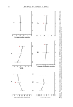
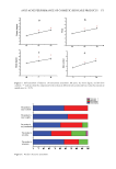
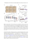
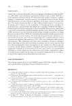
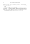
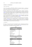
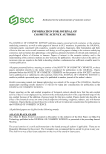
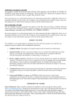
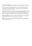
586 JOURNAL OF COSMETIC SCIENCE MEASUREMENTS TEWL was measured using a Tewameter TM-300 (Courage+Khazaka electronic GmbH, Germany). The results were expressed in g/m2/h. Skin moisture was assessed by measuring an electrical capacitance of the skin using a Corneometer CM825 (Courage+Khazaka electronic GmbH, Germany) in the laboratory. Moisture content was displayed digitally in arbitrary units (A.U.). Subjects self-recorded the degree of itchiness on the visual analog scale, ranging from 0 to 10 (No itching: 0 Mild itching: 1–less than 3 Moderate frequent itching: 3–less than 7 Severe itching: 7–less than 9 Very severe itching: 9–10) over the past 24 h at each institution visit on days 0, 14, and 28 days. STATISTICAL ANALYSIS Data are presented as the mean ± standard deviation (n = 3 for qPCR and ELISA data) or mean ± standard error of the mean (n = 33 for clinical trial data). Statistical analysis was performed using one-way analysis of variance followed by Dunnett’s or Sidak’s multiple comparisons test using GraphPad Prism (version 9.2.0 GraphPad Software, San Diego, CA, USA). For all analyses, differences with p 0.05 were considered statistically significant. RESULTS AND DISCUSSION ANALYTICAL HPLC PROFILES We performed HPLC analysis using an Agilent 1260 Infinity system to characterize the phytochemical components of the ethanolic extracts. We used precocene II, saikosaponin A, and gomisin A/gomisin N/angeloylgomisin H as reference compounds in A houstonianum, B falcatum, and S chinensis, respectively. The chromatograms of A houstonianum, B falcatum, and S chinensis were compared by the retention time to that of the reference standard compound (Figure 1). These data suggest that A houstonianum, B falcatum, and S chinensis contained active phytochemical compounds. ETHANOLIC EXTRACTS OF THE THREE DIFFERENT HERBS DO NOT INDUCE HACAT CYTOTOXICITY First, we determined the cytotoxicity of the three herbal ethanolic extracts. HaCaT cells were treated with different concentrations (0–80 μg/mL) for 24 h, and a cell viability assay was performed. Treatment with A houstonianum, B falcatum, or S chinensis did not induce considerable cytotoxicity up to 80 μg/mL compared to the vehicle-treated control (0 μg/mL) (Figure 2A). A combination of the three extracts showed no significant cytotoxicity until at least 48 h after treatment (Figure 2B). In the following experiments, we routinely used the combination of 40 μg/mL A houstonianum, 20 μg/mL B falcatum, and 40 μg/mL S chinensis. THREE HERBAL EXTRACTS RECOVER FLG EXPRESSION REDUCED BY IL4 + IL13 STIMULATION AD is one of the most common chronic inflammatory skin diseases characterized by prolonged and intense itching (34), making it ideal for studying the pathophysiology of pruritus. The well-known genetic risk factor for AD is a null mutation in the gene encoding

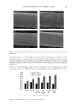
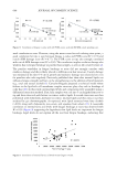
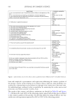
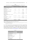
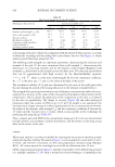
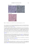
587 SUPPRESSION OF ITCHING BY THREE HERBAL ETHANOLIC EXTRACTS Figure 2. Effect of the ethanolic extracts of three selected herbs on cytotoxicity in HaCaT cells. HaCaT cells were treated with different concentrations (0–80 μg/mL) of A houstonianum, B falcatum, and S chinensis for 24 h (A) or a combination (40 μg/mL A houstonianum, 20 μg/mL B falcatum, and 40 μg/mL S chinensis) for varying lengths of time (0–48 h) (B). Cell viability was measured using a commercial water-soluble formazan-based assay kit (Cell Counting Kit-8). Figure 1. Analytical HPLC profiles of authentic standard and ethanolic extracts. Chromatograms of ethanolic extract of A houstonium (A), ethanolic extract of B falcatum (B), and ethanolic extract of S chinensis (C). Profiles of authentic standard compounds (STD 0.1 mg/mL) were plotted in the upper panel.
Purchased for the exclusive use of nofirst nolast (unknown)
From: SCC Media Library & Resource Center (library.scconline.org)