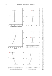
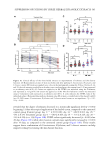
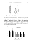
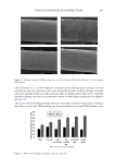
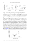
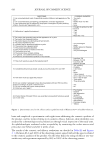
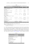
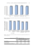
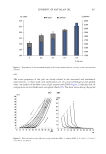
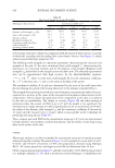
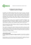
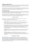
592 JOURNAL OF COSMETIC SCIENCE (Figure 8B), and immunoblot analysis (Figure 8C), respectively. Under these experimental conditions, we examined whether A houstonianum, B falcatum, or S chinensis affected IL4- induced TSLP expression in HaCaT keratinocytes. RT-PCR (Figure 8D) and qPCR (Figure 8E) showed that treatment with A houstonianum, B falcatum, and S chinensis alone or in combination significantly (p 0.001) reduced IL4-induced TSLP mRNA expression. Similarly, the IL4-induced increase in TSLP protein levels was significantly (p 0.01) reduced by single or combination treatment (Figure 8F), where the latter was more effective than the former. From these results, it is clear that A houstonianum, B falcatum, and S chinensis can inhibit IL4-induced pruritogenic TSLP production, and a combination of the three is more effective than each alone. THE THREE HERBAL EXTRACTS INHIBIT IL4–INDUCED POMC EXPRESSION POMC is a precursor polypeptide that generates numerous functional peptide hormones, such as adrenocorticotrophic hormone, melanocyte-stimulating hormone, and β-endorphin (53). β-endorphin, similar to morphine, is an endogenous opioid neuropeptide associated with relieving stress and depression (54). However, keratinocyte-derived β-endorphin stimulates sensory nerves in the skin, leading to itching behavior (e.g., β-endorphin is highly expressed in skin lesions, including AD, allergic contact dermatitis, and psoriasis) (55,56). β-Endorphin is produced via cleavage of the C-terminal region of the precursor POMC protein (57). We observed time-dependent induction of POMC mRNA expression after IL4 stimulation in HaCaT cells using RT-PCR (Figure 9A) and qPCR (Figure 9B). In Figure 7. Fluorescent immunocytochemical staining of IL31 in HaCaT cells stimulated with IL4 in the absence or presence of the three herbal extracts. HaCaT cells cultured on coverslips were treated with IL4 (20 ng/mL) in the absence or presence of the combination of the three herbal extracts (40 μg/mL A houstonianum, 20 μg/mL B falcatum, and 40 μg/mL S chinensis) for 36 h. After fixing and permeabilization, immunofluorescence staining was performed using anti-IL31 primary and Alexa Fluor 555-conjugated secondary antibodies (red fluorescence). Tubulin was counterstained using antitubulin primary and Alexa Fluor 488-conjugated secondary antibodies (green fluorescence). Nuclear DNA was visualized with Hoechst 33258 (blue fluorescence). The areas in the dashed boxes are magnified in the right panels. Scale bars, 400 μm.

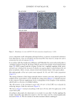
593 SUPPRESSION OF ITCHING BY THREE HERBAL ETHANOLIC EXTRACTS addition, the abundance of secreted β-endorphin in the culture medium was also increased significantly (p 0.01) after 24 h of IL4 stimulation (Figure 9C). Next, we determined whether A houstonianum, B falcatum, or S chinensis affected IL4-induced POMC expression in HaCaT keratinocytes. RT-PCR (Figure 9D), qPCR (Figure 9E), and immunoblot analysis (Figure 9F) showed that pretreatment with A houstonianum, B falcatum, S chinensis, or a combination of the three significantly (p 0.01) reduced IL4-induced POMC expression. Furthermore, ELISA revealed that IL4-induced accumulation of secreted β-endorphin was also significantly (p 0.001) reduced in the culture medium by treatment with A houstonianum, B falcatum, S chinensis, or a combination of the three (Figure 9G). These data demonstrate that A houstonianum, B falcatum, or S chinensis inhibit IL4-induced pruritogenic Figure 8. Effect of the three herbal extracts on the suppression of IL4-induced TSLP expression in HaCaT cells (A and B). HaCaT cells were treated with IL4 (20 ng/mL) for various time periods (0–36 h). Total RNA was isolated, and TSLP mRNA levels were examined by RT-PCR (A) and qPCR (B). GAPDH was used as the loading control (C). HaCaT cells were treated as in (B), and whole-cell lysates were immunoblotted using anti- TSLP antibodies. GAPDH was used as the loading control. Band intensities were measured using Image J (D and E). HaCaT cells were treated with IL4 (20 ng/mL) for 12 h in the absence or presence of A houstonianum (40 μg/mL), B falcatum (20 μg/mL), S chinensis (40 μg/mL), or a combination of the three. Total RNA was isolated, and IL31 mRNA levels were examined using RT-PCR (D) and qPCR (E). GAPDH was used as the loading control (F). HaCaT cells were treated as (E), and whole-cell lysates were immunoblotted using anti- TSLP antibodies. GAPDH was used as the loading control. Relative band intensities were measured using ImageJ. ns: not significant *p 0.05, **p 0.01, ***p 0.001 compared to control (n = 3).
Purchased for the exclusive use of nofirst nolast (unknown)
From: SCC Media Library & Resource Center (library.scconline.org)