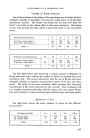
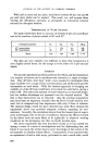
THE SKIN AS A COMMUNITY OF STRUCTURES E. W. POWELL, 1VI.A., A.R.I.C.* In l•art II of his article the author discusses the formation ot • skin l•igments and ot • hair. These two l•rocesses are carried out by cells of •wo distinct ty10es which respond ditterently to some topical influences, but are linked by certain common nutritional needs. PART II PIGMENT FORMATION AND KERATIN PRODUCTION IN THE SKIN THE PIGMENTS characteristic of the mammalian skin and its associated structures are the melanins, complex polymeric materials giving black, brown and red colours as well as providing the dark absorbing background for certain blues. It is now generally accepted that these materials are formed by the activity of certain specialised cells, the melanocytes. In or close to the basal layers of the epidermis and about the proliferating layer of the hair bulb are situated cells possessing a main body and branches. These are the pigment-forming melanocytes. In the skin, the two activities of keratin production and of the formation of pigment are, therefore, carried out by cells of two distinct types and of differing origins. ORIGIN AND DEVELOPMENT OF PIGMENT-PRODUCING CELLS During a relatively short period in the growth of the mammalian embryo there is associated with the rudimentary spinal cord a small amount of a tissue termed the neural crest. This is a transitory structure, for the constitu- ent cells disperse and move to positions where they lie at the interface between an epithelium and an underlying tissue. Such migration results in a fairly even distribution of these cells through the epidermis of the human fcetus, and when a hair follicle begins to develop they move down into the dermis with the descending follicle (Zimmerman, 1953). None of these former neural crest cells appear to remain in the upper part of the folhcle, and it is of interest that when sweat gland rudiments are formed none of these cells move down from the epidermis with them. In later development, these cells which have moved into the epidermis and hair bulbs can assume the branched form typical of nerve cells. The branches of these cells pass between the epidermal cells and end on them. When active, they produce a substance, dihydroxyphenylalanine, which is related to the noradrenalin formed in other nerve cells, and it is this substance which undergoes oxidation and poly- merisation during the formation of melanin. The melanin is passed along the branches of the melanocytes and is deposited between and also enters the epidermal cells. * 49, Barn Hill, Wembley Park, Middx. 276

THE SKIN AS A COMMUNITY OF STRUCTURES The origin of the melanocytes of the mouse has been beautifully demon- strated by Rawles. Grafts of the presumptive epidermis and dermis of embryo mice were transferred to a suitable part of an incubating chicken's egg when development proceeded in association with the chick (RaMes, 1953). The eggs were those of the White Leghorn and the tissues of the chick develop no melanin pigmentation. The grafts were taken from embryos before and after migration of the neural crest and in addition neural crest material itself was cultivated. The mice used were of a strain which de- veloped black hair, but formed no melanin in the skin. Under the conditions of cultivation, dermis and epidermis with hairs were developed normally, but grafts removed before migration of the neural crest grew white hairs only. Those removed after migration gave rise to black hairs. Moreover, the cultivated neural crest cells were found to migrate in the epithelia of the chick host and assume their normal form and pigmentary activity. There are many questions remaining to be answered concerning the factors controlling the migration and distribution of the prospective melanocytes, and also concerning their subsequent behaviour. It seems established that these cells undergo growth and division as do other cells, and also that they are included in the outward movement and shedding of the epidermis. Likewise they move outward from the hair bulb in the growth of the hair. During the period of rest of the hair bulb in which a hair club is formed the fate of the melanocytes in that bulb is not known precisely. Later, when the follicle and papilla become active and the complete structure is recon- stituted, it is likely that the melanocytes which pigment the new hair arise from precursors in the residue of the previously active hair bulb (Chase et al.). The presence of melanocytes in tissue can be demonstrated by certain staining techniques, and such methods have shown that there are similar numbers per unit area in white and negro human skin. The difference in racial colour in this and other instances is due to differences in the activity of the cells and is genetically controlled. In the condition of albinism, melanocytes are present in the skin, but remain incapable of forming melanin. Difference in activity as shown by the colour of the melanin pro- duced is responsible for the colour pattern in certain guinea-pigs which possess numbers of melanocytes in red areas of skin similar to those in black areas (Billingham & Medawar, 1953). In common with other living tissues, the pigment-producing cells function through the activity of bio-catalysts, the enzymes. One enzyme associated particularly with the formation of melanin is tyrosinase, which can convert the amino acid tyrosine to dihydroxyphenyla!anine, the precursor of melanin. Although emphasis is placed on the particular chemical reactions involving the oxidation and polymerisation of these single amino acids, tyrosine and dihydroxyphenylalanine, melanin is associated with protein in the living 277
Purchased for the exclusive use of nofirst nolast (unknown)
From: SCC Media Library & Resource Center (library.scconline.org)