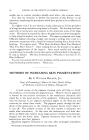
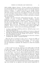
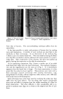
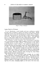
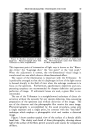
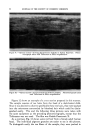
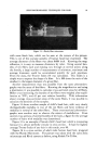
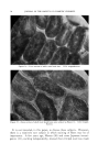
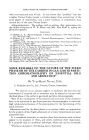
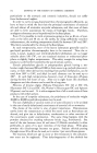
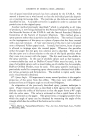
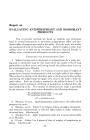
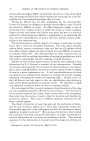
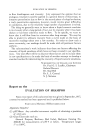
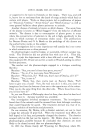
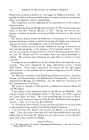
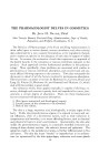
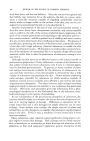
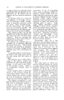
PHOTOMICROGRAPHIC TECHNIQUES FOR HAIR 91 Figure 7.--"Optical section" showing continuous medulla in human head hair. Photomicro- graph taken with Trilineator at 500X magnification. .. "•.:-•? ?•--.,... '-:..::.,, . - ............. .,::.,- ß ..... •---•. .-.• ....... ' ........ •"s-- ..x.....?- :• .... ---..-'.•,--...-.:F,':.?/?5-. ..... .x•:• '. : :•:•* •' - ..... .% -,' •- :*:: :,,v': '"'*' :'"." ...... . .... •- .- r-. "r '** """ : • '*'. .... Figure 8.•-"Optical section" showing discontinuous medulla in human he ad hair. Photo- •- micrograph taken with Trilineator at $00X magnification. terial, such as paraffin or some resin or polymer, and then slicing sections with a rotary microtome. These methods are not suitable for human hair because the fibers slip too easily out of the embedding material when the blade hits them. Accordingly, a variety of special microtomes have been devised for fibrous materials, and a description of them can be found in any of the textbooks on fiber microscopy. The simplest of these, and one which is quite suitable for work with human hair, is the Hardy Fiber Mi- crotome, which is shown in Fig. 11. To use this device, the two halves are disengaged, and a bundle of fiberõ' is inserted in the slot and held in pl&ce by inserting the tongue of the other half. The sample is'cut off flush with both faces of the plate, and the desired thickness of section is pushed out through the slot by means of a graduated micrometer screw •rrangement. Before slicing off the section" with a razor blade, the speci- men is cemented together with collodion. "'

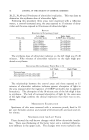
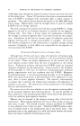
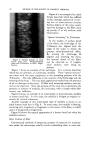
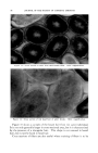
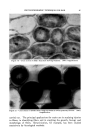
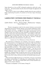
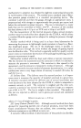
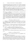
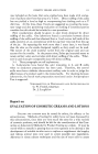
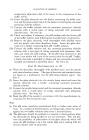
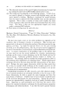
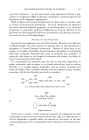
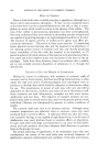
92 JOURNAL OF THE SOCIETY OF COSMETIC CHEMISTS Figure 9.--"Optical section" showing discontinuous medulla in human head hair. Photo- micrograph taken with Trilineator at 500X magnification. Figure 10.--"Optlcal section" of human head hair without medulla. Photomicrograph taken with Trilineator at 500X magnification. Figure 12 shows an example of a cross section prepared in this manner. The sample consists of ten hairs from the head of a dark-haired child. Since it was desired to observe specifically these ten hairs, they were packed into the microtome surrounded by bleached hair which could be distin- guished easily. This and the following three pictures were taken with the same equipment as the preceding photomicrographs, except that the Trilineator was not used. The film was Kodak Panatomic X. As a contrast, Fig. 13 shows some red hair from a female adult human head. The individual pigment granules are easier to see in this picture. To distinguish easily the ten fibers of the sample, they were packed in
Purchased for the exclusive use of nofirst nolast (unknown)
From: SCC Media Library & Resource Center (library.scconline.org)