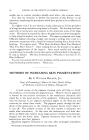
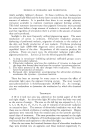
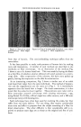
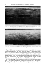
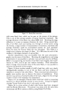
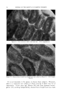
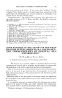
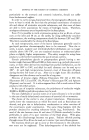
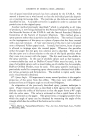
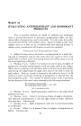
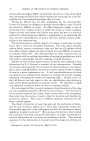
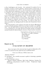
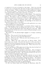
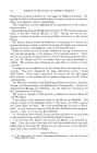
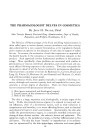
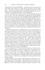
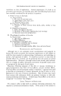
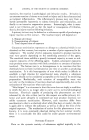
PHOTOMICROGRAPHIC TECHNIQUES FOR HAIR 87 Figure 1.--Cast of an adult male head hair. 330X magni- fication. Figure 2.--Cast of an adult male beard hair, same subject as Figure 1. 330X magnification. from that of keratin. The semi-embedding technique suffers from the same defect. It has been possible to study scale patterns of human hair by making casts and impressions. A number of such methods are described in the textbooks on fiber microscopy. As an illustration of the method, Fig. 1 shows a cast of a human head hair. This was made by laying the fibers on a thin film of collodion solution (diluted with amyl acetate) on a micro- scope slide. After evaporation of the solvents, the hairs were pulled up gently, leaving the impression on the slide for examination. As an interesting comparison, Fig. 2 shows a similar cast of an adult male beard hair taken from the same individual. The method of prepara- tion and the magnification are the same as in Fig. 1. It is immediately apparent that the beard hair is larger. On closer examination, it is also noted that the scales lie closer together. These pictures were taken with a Zeiss monocular research microscope with rotating micrometer s age, using Kodak Tri-X film, a Rival single lens reflex camera, and a 100-watt microscope lamp with focusing lens. Such techniques have often been used for studying the scales, but they suffer from two basic defects. For one thing, they require considerably more time and effort to prepare than the simple mounting of a fiber for direct observation. The other difficulty is that considerable skill is re- quired. to pr9duce ca•s.ts •:•[ are as sharp as the original and free of arti- facts, " , • .

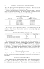
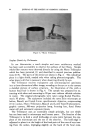
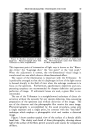
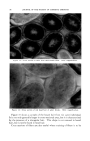
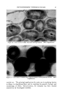
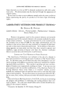
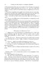
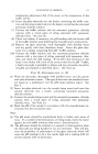
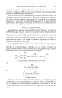
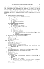
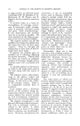
JOURNAL OF THE SOCIETY OF COSMETIC CHEMISTS Figure 3.--Munn Trilineator. Surface Details by Trilineator In our laboratories a much simpler and more satisfactory method has been used successfully to observe the surfaces of hair fibers. Simple whole fiber mounts were observed directly by using the Munn Trilineator which has been patented (5) and described in several previous publica- tions (6-8). The parts of the device are shown in Fig. 3. The cylindrical piece is a light shield, needed only when taking photomicrographs. The other piece is all that is necessary when observing directly by eye. The Trilineator resolves transparent and semitransparent specimens into a three-dimensional effect image, revealing to the eye or to the camera a detailed picture of surface structure. An illustration of this with a human head hair is shown in Fig. 4. The sample was prepared by ex- tracting with ether and mounting in 20 per cent calcium chloride solution in water. The original photographs were taken using Kodak Panchro- matic M plates. The equipment included the same Zeiss microscope as before, Bausch and Lomb 8-mm. apochromatic objective, compensating (15X) ocular, Munn Trilineator, Bausch and Lomb Type H photomicro- graphic camera, 500-watt projection lamp, focusing lens, fused Pyrex water cell and automatic exposure timers. The Trilineator uses an essentially simple principle, but one which has not been used in microscopy until recent years. The function of the Trilineator is to hold a small knife-edge at some point between the eye- piece of the microscope and the eye of the observer. The knife-edge is adjusted to place it at the height of the focal point of the cone of rays issu- ing from the ocular, impinging slightly on the back of the focal cone.
Purchased for the exclusive use of nofirst nolast (unknown)
From: SCC Media Library & Resource Center (library.scconline.org)