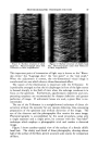
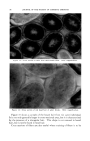
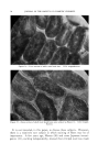
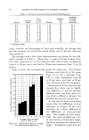
82 JOURNAL OF THE SOCIETY OF COSMETIC CHEMISTS 20, 25, 30, 40 and 50 minutes of ultraviolet irradiation. This was done to ddtermine the erythema dose of ultraviolet light. Following this procedure three areas were examined with a reflection meter a control untreated area, the area exposed to 15 minutes of ultra- violet and the area exposed to 50 minutes of ultraviolet light. RESULTS REaCT•O• TO ULTRaV•OLEX L•C•T Left thigh, Right thigh, Time, min. ultraviolet alone 8-MOP plus ultraviolet 15 No erythema No erythema 20 No erythema No erythema 25 Faint erythema No erythema 30 Definite erythema No erythema 40 Definite erythema No erythema 50 Definite erythema No erythema The erythema dose of ultraviolet radiation on the left thigh was 25-30 minutes. Fifty minutes of ultraviolet radiation on the right thigh pro- duced no erythema. REVLECT•O• METER REim•C WroTE D•sc = 50 Left Thigh Right Thigh Untreated area 33 31 Area exposed to 15 min. ultraviolet 30.5 26 Area exposed to 50 min. ultraviolet 27 28 (erythema) The relationship between the control areas and those exposed to 15 minutes of ultraviolet radiation indicates greater absorption of light in the area exposed after the ingestion of 8-MOP (probably due to pigment formation). The absorption of the 50-minute area of the left thigh is due to erythema. The lack of increased absorption of light by the same area of the right thigh confirms the clinical impression that there was no ery- thema. MICROSCOPIC EXAMINATION Specimens of skin were removed with a cutaneous punch, fixed in 10 per cent formalin solution and stained with hematoxylin and eosin and sil- ver nitrate. SPECIMENS FROM THE LEFT THIGH These showed the well-known changes which follow ultraviolet irradia- tion. There was thickening of the horny layer and a minimal inflamma- tory infiltrate in the upper curls. The changes were more marked in the

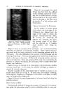
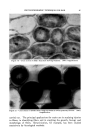
METHODS OF INCREASING SKIN PIGMENTATION 83 specimen removed after 14 days. Silver nitrate stain revealed almost no new pigment formation. SPECIMENS FROM THE RIGHT THIGH The horny layer was thickened and a specimen removed after 14 days of ultraviolet irradiation showed the formation of a stratum lucidum. This is a homogenous pink staining layer lying at the lowest level of the stratum comeurn. Normally this is present only in the skin of the palms and soles. The cells of the epidermis were swollen and there was inter- cellular edema. The perivascular infiltrate in the upper cutis was more prominent than that seen in specimens from the left thigh. Silver nitrate stain of the specimen removed after 14 days of irradiation revealed new pigment formation in the melanocytes. The amount of melanin, however, was not very great. No pigment was present in the horny layer. In order to determine the effect of 8-MOP over a longer period of time, a biopsy specimen was removed from the back of a patient who had been using 8-MOP and ultraviolet light for the treatment of vitiligo for two months. His skin was darker than his usual tan. This specimen had a very prominent stratum lucidum. In spite of this individual's dark tan the histologic sections did not reveal abnormally active pigment formation. Most of the pigment was contained in cells in the basal cell layer (prob- ably the cell bodies of melanocytes). A few dendrites contained pigment granules and there were irregularly scattered pigment granules in the horny layer. Although a controlled experiment was not performed on this patient, the amount of pigment retained by the melanocytes was much greater than one would expect in this individual (light complected, blue eyed Scandinavian). INTERPRETATION The initial effect 'of 8-MOP plus ultraviolet radiation occurs in the upper layers of the epidermis. This is as far as the high energy, erythema- producing ultraviolet radiation penetrates. The metabolism of the cells is altered in such a way that a stratum lucidurn is formed. This struc- ture explains the increased tolerance to ultraviolet light (increase in ery- thema dose) which occurs before there is any appreciable pigment forma- tion. Taking 8-MOP, increased pigment formation. There was also an in- crease in the inflammatory reaction and pigmentation did not precede the inflammation. These findings appear to rule out a direct effect of 8-MOP alone or 8-MOP and high energy ultraviolet radiation on the pigment form- ing system. We cannot be certain about the effect of low energy, pene- trating ultraviolet. It is fairly certain that long wave ultraviolet and
Purchased for the exclusive use of nofirst nolast (unknown)
From: SCC Media Library & Resource Center (library.scconline.org)