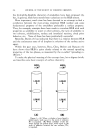
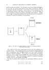
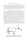
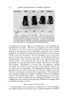
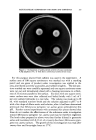
MAINTENANCE OF SKIN IN VITRO AS AN ORGAN SYSTEM 343 Our research indicated that there were two salient limiting factors present in maintaining organ cultures of adult skin. These were (1) the diffi- culty of supplying adequate nourishment to the multilayered tissue by dif- fusion alone and (2) the process of cellular dedifferentiation which occurs in vitro, with subsequent deterioration of the architecture of the skin. In a series of experiments using both animal and human skin a technique w/rs developed by which we are largely able to circumvent these problems. Adult skin has been successfully maintained in vitro for as long as four weeks. MATERIALS AND METHODS The initial technical problem was to obtain full-thickness skin which would be thin enough to permit adequate diffusion of nutrients to the epidermis. In the series of experiments in animals the ears of adult mice were chosen as donor sites. By using the auricular skin as our experimental tissue we are able to obtain full-thickness grafts, with the epidermal, derreal, and fatty layers intact, which are extremely thin and are large enough to be followed after grafting, i.e., around 20 ram. 2 in area. Also, when transplanted to the dorsum of a mouse the auricular skin retains its characteristic appearance and can always be easily grossly identified. The technique used to obtain the auricular skin is our adaptation (5) of a method originated by Edgerton (6). Immediately after excision, the graft is soaked for one-half hour in a Petri dish containing a suspension of 200 units/cc. of Mycostatin in Earle's Balanced Saline solution. The graft is positioned in the suspension with the epithelial surface downward on a piece of gauze. Next, the graft is placed for one hour on gauze saturated with a solution of 2000 unit/cc. crystalline penicillin potassium and 250 mg/cc. Streptomycin in Earle's Saline. Care is taken to spread the graft evenly over the gauze, with the epithelial surface upward. Before the grafts are placed in culture flasks, 0.1 cc. of a •25 mg/cc. aqueous suspension of Cortisone Acetate is introduced with a tuberculin syringe into each flask. The flask is held tilted so that the liquid streaks down one side of the vessel. The water is evaporated from the suspension by gently flaming the flask and an even film of cortisone remains adherent to the vessel wall. The cortisone is added to inhibit the differentiation and the subsequent migration of the fibroblasts from the dermis (7). Each graft is then transferred with long fine pointed forceps, to the sterile 4.5 culture flask and evenly spread out over the bottom surface. The addition of the culture medium detaches the graft from the glass surface so that it floats freely, with the epithelial surface upward and the dermal layer in contact with the medium. The culture medium determined to be adequate through experimenta- tion (8), consists of Eagle's Medium with a 10 per cent supplement of

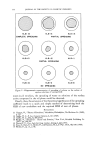
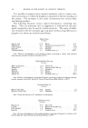
344 JOURNAL OF THE SOCIETY OF COSMETIC CHEMISTS horse serum, a 1:100 dilution of a standard penicillin-streptomycin solution for tissue culture and a 100 unit/cc. suspension of Mycostatin. The medium for cultures that are to be maintained for two weeks or longer contains an additional 5 per cent supplement of beef embryo extract and a 10 per cent supplement of ascitic fluid (9). These constituents are all a[ailable from Microbiological Associates. Each graft receives 4 cc. of fresh medium three times a week. Care must be exercised to remove and renew the medium without wrinkling the skin, since optimum diffusion of nutrients through the tissue is desired. After ten days in culture, 0.1 cc. of the cortisone suspension is added directly to the fluid medium. In the experiments utilizing human skin a similar technique is employed. The donor site, however, depends on the available patients who volunteer. It is usually the thigh. The area is depilated and prepared with PHiso- hex* and 70 per cent ethanol. The skin is removed in the operating room by routine surgical procedure. Generally, strips of skin 2 cm. wide and 12 cm. in length are cut at 18 thousandths of an inch. Epidermis and some dermis is obtained although the grafts are not full thickness. The strips are then divided into 6 grafts, each 2 cm? in area. The grafts are transported to the laboratory in sterile containers and prepared for tissue culture as described for the mouse auricular skin. Large culture flasks roughly 200 cc. in volume are used. One cubic centimeter of the cortisone suspension is added to each vessel and flame dried. Twenty-five cubic centimeters of the described medium is inserted under each graft and renewed once a week. In every series some grafts were sacrificed for histologic examination. The criterion in our study for the maintenance of the structural integrity of the auricular skin is the subsequent successful grafting of the skin to the original donor. The grafting procedure is described in the literature (10). The human skin was homografted to voluntary recipients and the course of these grafts was compared to that of skin homografted directly, without prior residence in tissue culture. RESULTS Basic studies to determine the nutritional requirements of adult skin in vitro have been completed. In a series of preliminary studies, 66 grafts of full-thickness auricular skin of mice were maintained in different media containing various nutritional supplements without the addition of corti- sone. A complex medium containing serum proteins proved to be essential for prolonged maintenance of the grafts in vitro. Intact auricular grafts were successfully maintained in this medium for two weeks using the flota- tion technique developed in this laboratory. Of 30 auricular grafts cultured for three weeks in this medium 16 per cent survived. At the end of three * 3.0 per cent solution of hexachlorophene in PHisoderm.
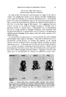
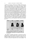
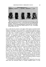
Purchased for the exclusive use of nofirst nolast (unknown)
From: SCC Media Library & Resource Center (library.scconline.org)