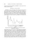
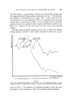
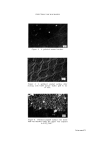
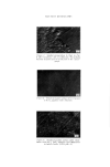
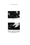
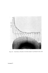
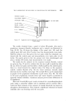
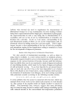
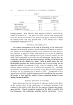
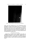
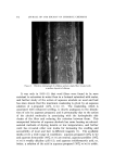
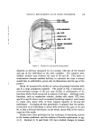
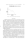
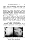
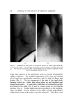
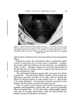
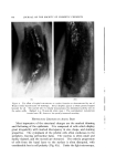
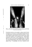
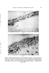
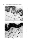
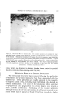
ELECTRON MICROGRAPHS Figure 10. Preparation shown in Fig..9 after brushing. Figure 11. A reflectance electron micrograph of an enamel surface. The area to the right was exposed to a 0. ! •o SnF2 solution, that to the left was masked. The complete tooth was then exposed to lactate buffer.

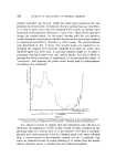
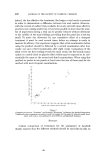
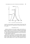
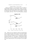
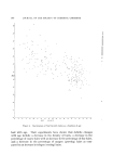
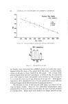
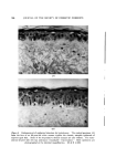
Figure 12. Distribution of fluoride in the labial enamel of a permanent incisor (14). Facing page 479
Purchased for the exclusive use of nofirst nolast (unknown)
From: SCC Media Library & Resource Center (library.scconline.org)