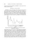
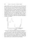
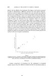
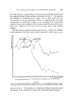
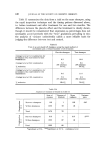
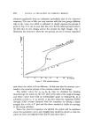
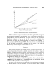
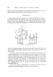
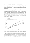
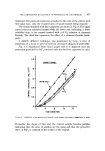
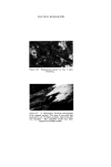
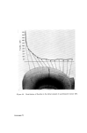
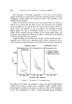
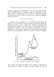
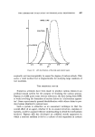
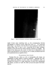
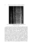
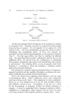
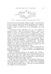
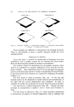
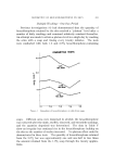
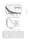
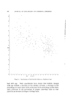
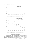
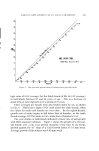
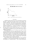
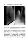
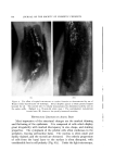
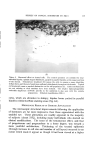
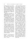
THE LABORATORY EVALUATION OF PROPHYLACTIC DENTIFRICES 481 solution as suggested by Brudevold (17). This is obviously more effective than when incorporated in a dentifrice though even then there is a sig- nificant uptake compared with the controls. The dotted line indicates the fluoride level above which the test teeth have acquired a significant increase in fluoride. TIN UPTAKE BY ENAMEL A similar type of experiment can be performed to demonstrate the presence of tin derived from stannous fluoride preparations though the analytical procedure is not so simple. The technique of electron probe microanalysis is applicable and the results can be obtained either as a photograph in which the atoms of tin appear as white spots on a dark ground or as a recorder trace obtained as the electron beam traverses the selected area. An example of the latter is shown in Fig. 14. A polished ENAMEL POLISHED • E NA.••/.,/- BACKGROUND Figure l•t. Tin uptake on polished and on etched enamel. tooth surface was etched with dilute acid to produce a white spot. A stannous fluoride treatment was given and, after rinsing, the area was

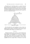
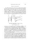
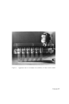
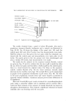
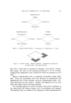
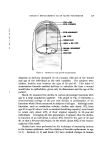
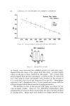
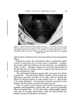
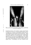
482 JOURNAL OF THE SOCIETY OF COSMETIC CHEMISTS scanned. The peaks on the trace indicate the presence of tin on the tooth surface and it can be seen that greatest uptake occurs in the white spot area. This may well be due to the increased surface available on this etched region rather than to any specific affinity of white spot enamel for tin. IN vivo METHODS All the tests mentioned are test-tube experiments and however much the conditions are varied in an effort to test the permanence of the treat- ment, they give no unequivocal indication of what will happen in the mouth. Waiter {18) was the first to describe a feasible technique which went some way to answering this criticism, in that during the period of the trial, the treated teeth were exposed to all the dynamic conditions normally present in the mouth. His test was again dependent upon solubility changes of tooth enamel but on humans it was conducted as follows: A 2mm diameter filter paper disc impregnated with an acid base indica- tor and with 2[d of 0.3N HC1, was placed on the selected tooth surface after it had been dried with a tissue. The time taken by the indicator to change colour {Colour Reaction Time) is a function of the tooth's 'resist- ance' to the acid attack. In a test using a panel of school children Mtihle- mann (19), using this method, reported the effectiveness of certain amine hydrofluorides. In our hands this test gave a positive result for a topical stannous fluoride treatment but even with half the amount of acid used by Mfihle- mann it had a (literally) marked effect on the subjects' teeth and had to be abandoned. An alternative method was then developed in which a weak acid was used in place of hydrochloric acid and the change in pH was recorded after a predetermined time. The technique is described by Middleton and Holmes (20) and Morley and Holmes (21), and depends upon clamping a well to an upper central incisor so that leakage cannot occur (Fig. 15). With the •vell clamped in position, 0.1 ml acetic acid (pH=3.4) is added from a syringe and is stirred by a stream of bubbles from the air line during the 3 min of the test. At the end of this time the air stream is stopped and a sample of the acid is sucked into the microcapillary glass electrode for pH measurement. This is a rather elaborate micro-electrode supplied by Messrs. Pye of Cambridge.
Purchased for the exclusive use of nofirst nolast (unknown)
From: SCC Media Library & Resource Center (library.scconline.org)