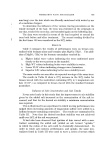
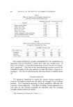
834 JOURNAL OF THE SOCIETY OF COSMETIC CHEMISTS seem to be, however, an unfortunate tendency to compact the phenom- enon of aging and attempt to have it fit one of these four theories to the exclusion of all others. From our viewpoint, it would seem that the theories mentioned here are actually very closely related. As has been shown, a mistake in the genetic material, the most basic yet most important controlling mechanism in the cell, might lead to inactive molecules which would be perfect candidates for creating a log jain in the cell. In addition, the wear and tear on an organism may be due to increased defects and inefficiency in cell function as such metabolic clogs and cross-linked compounds are formed. Metabolic by-products such as free radicals add to the impairment of the cellular operation. Thus, the problem of aging may be thought of as a steadily increasing cell disfunction at the molecular level, terminating in cell death. AGE-RELATED SKIN CHANGES After discussion of why externally manifest changes with age may occur, the next consideration should properly be the changes themselves. Some of the most readily apparent consequences of aging in the body are found in the area with which we are all so intimately concerned--the skin. In the following sections, then, the qualitative and quantitative age-related changes in the biochemistry and histology of the body's cutaneous covering will be examined. Derreal Changes: Collagen Most of the investigations to date into skin aging have centered on the dermis. Unfortunately, even as late as the first part of this decade, aging changes in the skin were being confused, and often equated, with changes brought about by exposure to ultraviolet light. Actually, it has been shown that specific differences exist between young and old skin, young and ultraviolet-exposed skin, and old and ultraviolet- exposed skin. Lorincz (10) has therefore suggested that the skin changes which were formerly classified as "senile elastosis" are actually a result of chronic ultraviolet exposure and should more properly be termed "solar elastosis." The epidermal and dermal layers of the skin represent, respectively, 5% and 95% by weight. The dermis contains 11% lipid, 1% carbo- hydrate, 7• nonfibrous protein, 79% collagen, and 2• elastin. Ob- viously, to quantitate any changes in normal or elastotic skin, the com- ponents believed to have changed must be separated from the skin. Sams and Smith (11) have described a representative procedure in which

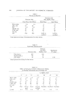
CHANGES IN HUMAN SKIN WITH AGING 835 the different fractions of collagen and elastin are separated by acid digestion and centrifugation. The collagen fractions are quantitated by measuring the amount of hydroxyproline in each fraction this amino acid is present in fairly constant amounts in the peptide linkages of each of the various types of collagen and, as such, is used as a marker for collagen. When the skin from the back or abdomen of a stillborn infant is compared with the skin from. the "Y" incision at autopsy of adult individuals, a decrease is found in the soluble fraction, and an increase in the insoluble fraction, of collagen from the adult skin in addition, there is an increase in total collagen per gram of wet weight tissue over the value for infant skin (11). That this is a true increase in collagen, and not just an apparent change due to water loss from the skin, is borne out by the findings of both Rothman (12) and Flesch (13) who reported an increase in the water content of senile skin as compared to infant skin. It should be noted that both of the above sites of tissue biopsy are not usually exposed to any great extent to the sun, so that the effects observed here are due to age alone. These changes could be explained by a phenomenon which was described earlier--namely, the cross linkage of collagen molecules. When the forearm skin of an aged donor is examined, it is found that there is no change in the soluble collagen fraction, but a remarkable decrease in the insoluble fraction, representing a decrease in total collagen over the value for the normal adult (11). These effects are commonly seen in the skin of the neck, forearm, face, etc., which has had chronic exposure to the ultraviolet rays of the sun this is usually referred to as sun-damaged skin. Smith and Finlayson (14) have advanced a theory which may explain these changes. They propose that collagenase is released from subcellular organelles located in the dermal fibroblasts called lysosomes when these lysosomes are labilized by ultraviolet rays below 3100 A. Therefore, upon repeated and chronic exposure to sunlight, the liberated collagenase digests and thereby reduces the dermal content of collagen. In addition to quantitative changes in dermal collagen with age, qualitative changes have also been recorded. Nimni et al. (15) have reported that there is an increase in the tensile strength of collagen with age, and that this increase in strength is in direct correlation with the known increase in insoluble collagen. A possible explanation for these results might again be the theory of cross linking between collagen molecules. In addition, Rasmussen et al. (16) found an age-
Purchased for the exclusive use of nofirst nolast (unknown)
From: SCC Media Library & Resource Center (library.scconline.org)