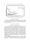
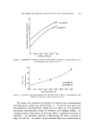
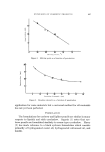
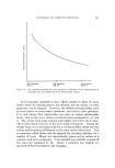
CHANGES IN HUMAN SKIN WITH AGING 837 like compound. However, Smith (22) and others have reported that this increase is due to true elastin build-up, basing their beliefs on the morphology, solubility, enzyme susceptibility, tinctorial and physical properties, and amino acid composition of the elastin formed. In quantitation of elastin from aged skin, diverse results are pre- sented. Sams and Smith (11), after chemical and physical separation of elastin from adult skin, concluded that there is a slight, quantitative increase with age. Weinstein and Boucek (23) subjected the elastin fraction to enzymatic digestion with elastase and found no increase in aged skin. Hult and Goltz (24) employed a procedure whereby they investigated the elastin content of human aged skin by two methods: the first, a microphotometric examination of orcein-stained skin sections, and the second, an enzymatic digestion. This was done to test whether there might be an increased susceptibility of elastin in aged skin to enzymatic digestion, rather than an increase in the quantity of elastin. They showed that there were indeed higher values for aged skin as opposed to young skin when the enzymatic method was used. The theory of age-related increased susceptibility might therefore appear to be true. But it would also seem that a positive answer has yet to be arrived at. EPIDERMAL CHANGES Turning now from the dermis to the epidermis of the skin, a first impression might be that the knowledge of the changes in protein and mucopolysaccharide content in the dermis with age has been gained at the expense of investigation of aging effects on the epidermis. Although some of the most apparent changes which accompany aging--skin wrinkles--are perceived in the outer layer of the skin, their true cause lies in the changes in dermal collagen and, possibly, elastin, which take place beneath the epidermis. Another common sign of aging is dry and flaking skin. At first this might seem hard to reconcile with the finding that there is an increased water content in human skin with increased age (13). However, it must be remembered that the outer horny layer of a healthy epidermis normally forms a barrier against the exchange of water, and that hydration of the skin surface is not influenced to any great extent by the water present in the deeper layers. Brief mention should also be made here of the effects that hormones can have on the aging skin. Although the role of the hormones in the body's homeostasis is all-encompassing, just one of the effects of the so-called sex hormones on the skin is presented here. In later life, and

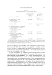
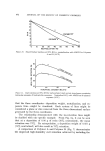
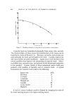
838 JOURNAL OF THE SOCIETY OF COSMETIC CHEMISTS especially in the female, who undergoes a rather abrupt decline in hormonal output at the menopause, the epidermal sweat and sebaceous glands become much less active, almost to the point of degeneration. Although the exact method of action of the sex hormone on the cell layers of the skin is not known, it is recognized that the "female" hormones and hormone-like steroids, when topically applied to aged skin, effect a histological picture in the epidermis which is strikingly similar to that of a young person. One area of investigation concerning the epidermis and aging that has been studied to some degree is that of rate of turnover of the cells of the epidermis. The turnover time, or renewal time, is usually defined as the average time for all the cells in the basal layer of the epidermis to reach the horny layer. Although several studies have been undertaken to measure this turnover time, various sources have been used for epidermal sections, and many different areas of the body have been used for tissue sampling. This lack of coordination between donor and site, coupled with infrequent recording of the donor's age, has unfortunately led to an unclear presentation of the relation of epidermal turnover to aging. Weinstein (25) has deduced values for turnover time using skin from the back of an adult pig. The morphology of the pig epidermis has been found to be strikingly similar to human epidermis. He found a transit time of 14 days through the viable cell layer, and 16 days through the horny layer, giving a total turnover time of 30 days. In working with the rat, Storey and Le Blond (26) reported a turnover time of 19 days in plantar epidermis taken from an adult animal. Bertalanffy (27), also using adult rats, found that the renewal time for the abdominal epidermis was 19 days. In a second study, Bertalanffy et al. (28) com- pared epidermal growth in young, adult, and senile rats. They found that when the total thickness of the epidermis from different age groups was compared, the senile skin showed a reduction by about two to three layers in the epidermis taken from ear or abdomen. In areas such as the bottom of the foot, there was an increase by one or two in the number of cell layers. As regards the mitotic rates, it was found that there was a general increase by about 50% in the rate of turnover of the senile epidermis as opposed to the young. In a similar study using human abdominal skin samples, Thuringer and Katzberg (29) observed an increase in the mitotic index such that the epidermis of the senile individuals was dividing twice as fast as that of the young group. The cause of this increased epidermal turnover is not yet positively
Purchased for the exclusive use of nofirst nolast (unknown)
From: SCC Media Library & Resource Center (library.scconline.org)