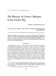
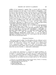
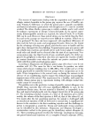
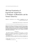
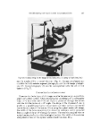
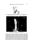
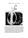
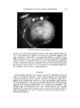
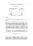
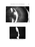
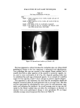
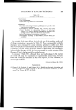
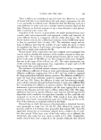
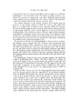
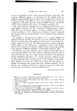
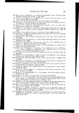
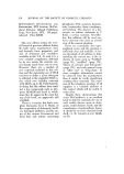
164 JOURNAL OF THE SOCIETY OF COSMETIC CHEMISTS accidentally be introduced into the eye, the cosmetics should be and are rou- tinely tested in animal eyes. The slit lamp becomes a tool which allows the investigator to make fine distinctions for ocular damage and between two or more comparative cosmetic materials. This laboratory is part of an ophthalmic pharmaceutical manufacturing company as such, the majority of the experimental and clinical research is in the ophthalmology area. Expertise has been developed in utilizing the slit lamp for the experimental ocular studies. Over the years, a grading system has been developed for toxicological and pharmacological evaluations of experi- mental studies in animals. This grading system depends upon the utilization of the slit-lamp biomicroscope as an indispensable experimental tool. There- fore, the gencral purpose of this and the next report is to present the uses and applications of the slit lamp in the experimental laboratory. The purpose of this endeavor is to describe the slit lamp and its use, and to illustrate the types of illumination employed. Figure 1. Slit • .... -..• :. i -51.. .•:• •:.• -: •::. . ß ..• : .... :•.: ....... •. '•.. -•$ . • .• .:: - ...... ......... •... . .• ..• ::: .... .•)•:• t-•': •..•? ......... •.'-... •$. : - •.. .-•. •..-• lamp with corneal microscope (a) •d s•t filuminator (b). (CouResy of Bausch and Lomb)

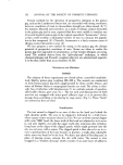
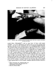
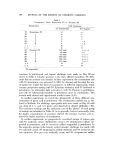
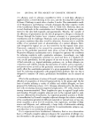
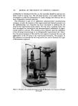
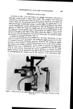
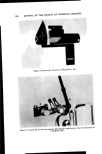
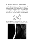
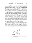
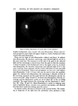
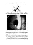
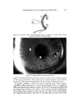
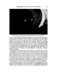
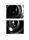
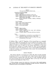
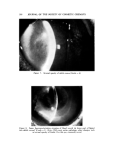
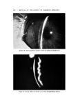
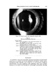
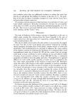
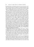
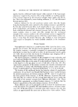
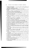
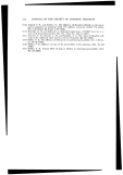
TECHNIQUES OF SLIT-LAMP ILLUMINATION 165 DESCRIPTION OF SLIT LAMP As shown in Fig. 1, the slit lamp is an upright instrument, which has a binocular eyepiece and a rather large ocular. The eyepiece and ocular are mounted on a movable ann so the slit lamp can be moved in a two-plane di- rection. There are two principal parts of a slit lamp. The optical part (a cot- heal microscope) includes the eyepiece and the different objectives for select- ing the magnification and the slit illuminator. The slit illuminator allows the operator to adjust the width, the height, and for lateral displacement of the slit image for certain specific types of illumination. In addition, filters can be placed in the path of the light so that either cobalt blue light or a green beam can be used. The slit illuminator and corneal microscope are situated to allow free rotation of each part through a 180 ø are. A number of attachments are available for a slit lamp. The Hruby (3) lens (Fig. 2) is used to view the fundus. The Goldmann applanometer (4, 5) (Fig. 2) is used to measure intraocular pressure. However, the Goldmann appla- riometer is not as useful in experimental animals as it is in the clinic the contour and wetting properties of the cornea of animals are apparentlylampbecausechamberusedtubebe different from man. A pachometer (6) can be mounted onto the slit (Fig. 3) and used for measuring the corneal thickness or anterior depth. Cameras can be mounted in the path of the slit image and can for photographing the images seen by the observer or an observation :'i ' ....... ' .............. .:7 '•:I.5 •--:' "-': ....-.,.b. ** :' ..'? *' ..................... * .... **.*'.•"i ...... .,.:.,.:. :.......::•-.. . : -?-" ...•::.- -%'•f•.•-.,---,-• .• :• ...•..•,• .•.• •:•. ..--:%.,.•.-"• '%* ............... • :'• ,' • • .' :.: , ..•:-..•.{ ...':' :• : , • :• •.. ...-? - • •-.:.::•.:"• ..:• •' ' .'• • -)C:":•* • ... .• . ...•. '.•c. ' .........
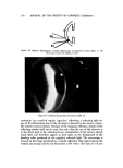
Figure "' **"'• -. 2. - :. "' ."• .:T-.•: ' .......: ........... * ' •. *-': *:, , 2. Hruby lens (a).d .planomet ' •-b,'" t "'•" •"':* **:"-. ::"•C:• .... Ze•s, In .) -• to s,it lamp. (Courtesy of Carl
Purchased for the exclusive use of nofirst nolast (unknown)
From: SCC Media Library & Resource Center (library.scconline.org)