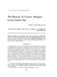
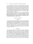
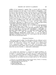
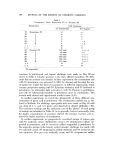
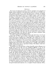
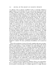
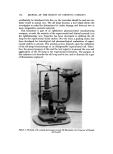
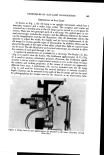
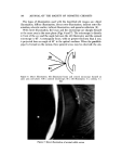
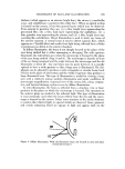
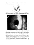
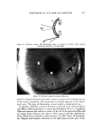
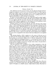
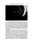
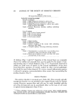
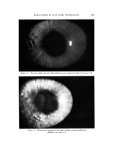
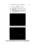
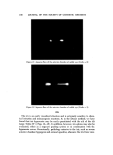
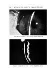
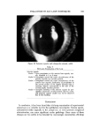
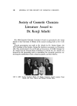
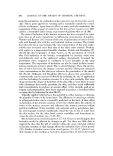
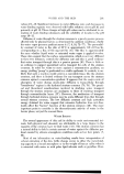
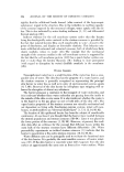
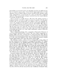
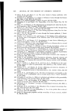
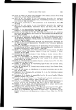
166 JOUBNAL OF THE SOCIETY OF COSMETIC CHEMISTS Figure 3. Pachometer. (Courtesy of Haig-Streit, Inc.) ' a' ":" ,•.. .... •:."• •, - •./$•. ii. •,_,":. • •. •': '"' • -.•_ .. -.....,-•&½'.•..' .s%•-.• ...... • .,,• . -. ?-••." - '•""• "- .... . ...- -'• ':-':4•' :..:• '•':•9•L• '" -: '-• .:. a.• •- -•6.. -'.'• 'a'*:.:":•'.:..' '.. :.:'•%.•. .. ß • : '• :• .. c, *. - '"' .' ' .•. '•% • .... , Camera (a) [or anterior segment photo•aphy and observer tube (b). (Conrtesoi Carl Zeiss, Inc.)

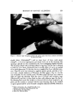
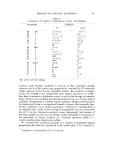
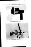
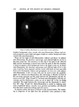
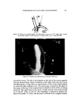
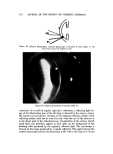
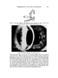
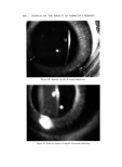
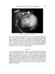
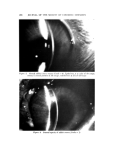
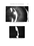
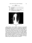
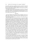
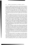
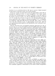
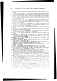
TECHNIQUES OF SLIT-LAMP ILLUMINATION 167 . . : Figure 5. Camera set-up for slit-image stereophotography. (Courtesy of Carl Zeiss, Inc.) may be employed for a second observer (Fig. 4). Camera attachments are available for both anterior segment photography and for slit image photogra- phy (7). Stereophotography (8) can be accomplished with the aid of two cameras (Fig. 5). TYPE OF SLIT-LA5IP ILLUI•{INATION There are txvo basic types of slit images used in biomicroscopy: a parallele- piped and optical section. When employing the parallelepiped, a rectangular beam (1-2 mm wide and 5-10 mm high) is projected through the cornea •vith the slit illuminator at a 45 ø angle. The shape of the illuminated area is similar to a parallelepiped prism whose outer and inner surfaces are curved due to the curvature of the cornea. When using the optical section slit image, the width of the beam is narrowed to almost its minimum and is projected from an angle of about 45 ø through the cornea. The result is a sagittal view or optical section similar to a thin histological section. The width of the anterior and posterior faces of the optical section should be about 20 ./z.
Purchased for the exclusive use of nofirst nolast (unknown)
From: SCC Media Library & Resource Center (library.scconline.org)