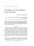
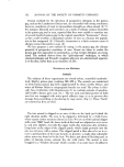
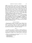
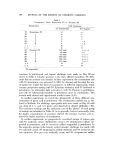
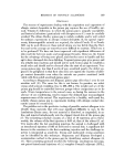
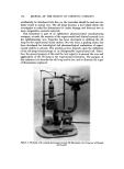
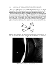
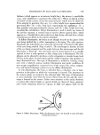
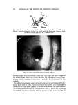
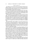
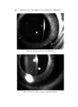
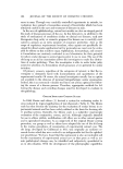
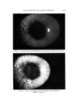
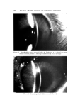
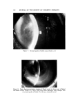
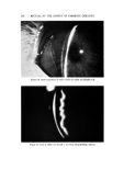
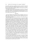
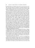
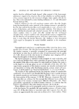
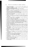
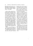
170 JOURNAL OF THE SOCIETY OF COSMETIC CHEMISTS Figure 9. Diffuse illumination of normal rabbit corneal epithelium brighter background when viewed with retro-illumination. Edema and cor- neal precipitates scatter light and appear bright on a darker background when viewed with retro-illumination. There are two types of retro-illumination: indirect and direct. In indirect retro-illumination, the structure, microscope, and reflected light do not lie in the same visual line. The structure is at the side of the path of the reflected light and the microscope is moved away from the path so that the structure is observed against a dark background such as the pupil or the iris. In direct retro-illumination, the structure, microscope, and reflected light all lie in the same visual line. Scars, pigment, and the vessels with blood are best seen with direct retro-illumination. Edema and precipitates are best seen with indirect retro-illumination. A slit 1-2 mm wide may be used to form the parallele- pipcd. For indirect retro-illumination, the microscope is directly in front of the eye being examined and the angle betxveen the illumh•ation and the ob- server is set at 45 ø (Figs. 10 and 11 ). In direct retro-illumination, both the mi- croscope and the illumination system are set at about 45* to the eye being examined. The angle between the microscope and the illuminator is, there- fore, 90 ø (Figs. 12 and 13). Retro-illumination is useful in studying most types of epithelial edema, vacuoles, scars, posterior precipitates, and channels from the blood vessels that infringe upon the cornea. In sclerotic scatter, the beam is focused on the corneal limbus. The scatter- ing or dispersion of the light from the perilimbal sclera produces a halo

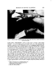
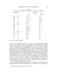
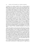
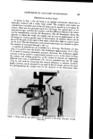
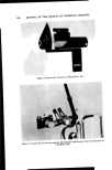
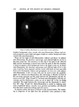
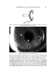
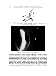
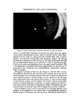
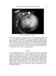
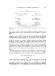
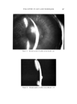
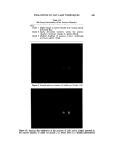
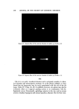
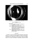
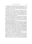
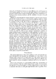
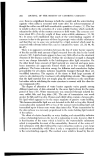
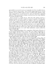
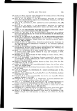
TECHNIQUES OF SLIT-LAMP ILLUMINATION 171 ß •.T. Figure 10. In'direct retro-illumination. Slit illuminator beam set at 45 ø angle with corneal microscope tangent to and focused on corneal surface. RL ----- reflected light Figure 11. Indirect retro-illumination of normal rabbit eye around the cornea. The halo is the brightest on the side of the cornea opposite h'om the focused beam. Internal reflection of the light transverses the cornea and illuminates it. Unless there are abnormalities of the cornea which impede or scatter this light, nothing is seen. The microscope is focused at the plane of the cornea in the area to be examined and is directly in h'ont of the eye being examined. The angle between the beam of light and the microscope should be at '_,east 45 ø (Figs. 14 and 15). The diaphragm is adjusted to produce the desirod slit width (1-2 mm) and the beam is focused at the temporal limbus.
Purchased for the exclusive use of nofirst nolast (unknown)
From: SCC Media Library & Resource Center (library.scconline.org)