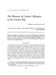
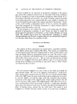
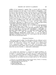
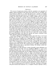
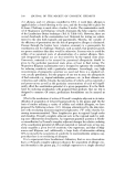
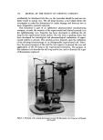
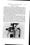
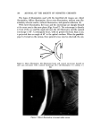
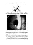
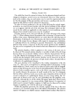
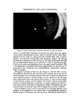
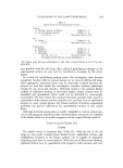
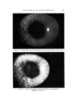
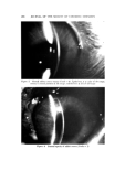
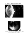
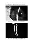
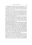
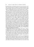
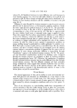
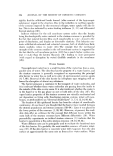
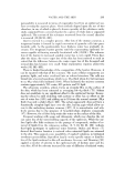
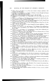
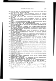
168 JOURNAL OF THE SOCIETY OF COSMETIC CHEMISTS The types of illumination used with the described slit images are: direct illumination, diffuse illumination, direct retro-illumination, indirect retro-illu- mination, sclerotic scatter, indirect illumination, and specular reflection (9). With direct illumination, the beam and the microscope are sharply focused at the same area in the same plane (Figs. 6 and 7). The microscope is directly in front of the eye and the angle between the slit illuminator and the corneal microscope is 45 ø. A rectangular beam, with no greater thickness than 2 mm, is projected from an angle of 45 ø to the optical medium. When the parallele- piped is formed on the cornea, three general areas may be observed: the epi- Figure 6. Direct illumination. Slit illuminator beam and cornea] microscope focused on same area and plane. CM--• corneal microscope SI----slit illuminator C--cornea I---- Figure 7. Direct illumination of normal rabbit cornea ... ..

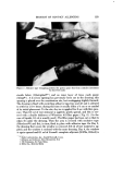
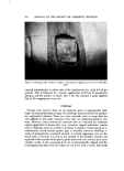
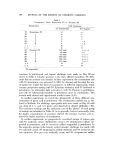
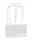
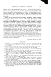
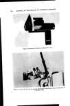
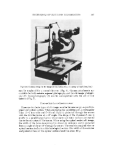
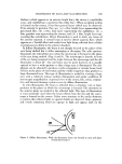
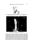
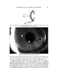
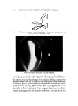
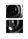
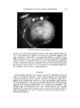
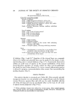
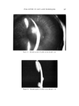
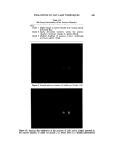
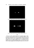
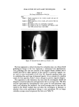
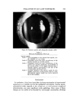
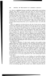
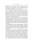
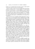
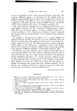
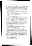
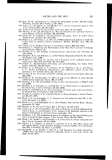
TECHNIQUES OF SLIT-LAMP ILLUMINATION 169 thelium (which appears as an anterior bright line), the st-roma (a marblelike area), and endothelium (a posterior thin white line). When an optical section is formed on the cornea, it has four general layers which may be observed. From anterior to posterior, they are: (a) a thin, bright layer representing the precorneal film (b) a thin, dark layer representing the epithelium (c) a thin, granular area representing the stroma and (d) a thin, bright layer rep- resenting the endothelium. Direct illumination is used to study any tissue of the anterior segment. A conical beam is used to detect aqueous flare, which appears as a Tyndall effect and results from light being reflected from cellular or proteinaceous debris in the anterior chamber. In diffuse illumination, the beam is not sharply focused in the plane of the area being studied but is either converging or diverging. The wide aperture illuminates the interorbital area when the microscope is focused in the plane of the area being studied (Figs. 8 and 9). The microscope is directly in front of the eye being examined and the angle between the microscope and the slit illuminator is about 45 ø . Any size beam may be used however, it is usually optimal to have a wide aperture so that a large area is illuminated. The dia- phragm can be adjusted to produce a wide rectangular or circular beam. Lack of focus in the plane of observation and the width of aperture thus produces a large illuminated area. This type of illumination is useful for viewing a large area with a relatively intense uniform illumination and under conditions of stereoscopic magnification. A general view of the cornea, lids, sclera, coniunc- riva, and lacrimal drainage system is seen by diffuse illumination. In retro-illumination, the beam is reflected from a structure (iris or lens) posterior to the plane on which the microscope is focused. The structures in the anterior plane are studied in the reflected light. This type of illumination is most commonly used when the beam reflects from the iris and the micro- scope is focused on the cornea. Unless some structure in the media obstructs or scatters this reflected light, no special details are observed. Scars, pigment, and vessels containing blood are opaque to light and appear dark on the Figure 8. Diffuse illumination. Wide slit illuminator beam not focused in area and plane corneal microscope is
Purchased for the exclusive use of nofirst nolast (unknown)
From: SCC Media Library & Resource Center (library.scconline.org)