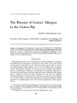
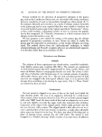
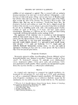
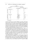
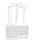
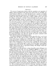
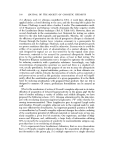
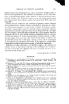
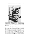
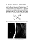
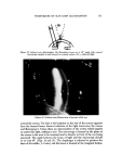
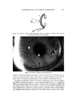
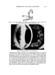
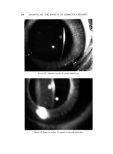
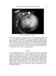
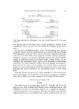
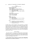
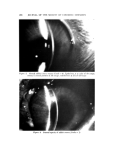
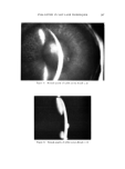
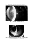
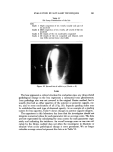
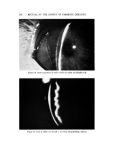
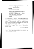
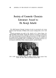
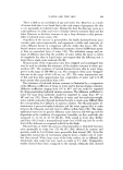
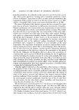
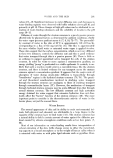
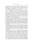
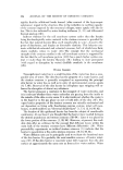
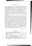
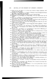
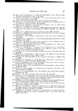
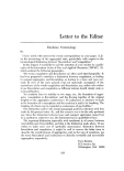
190 JOURNAL OF THE SOCIETY OF COSMETIC CHEMISTS Figure 11. Aqueous flare of the anterior chamber of rabbit eye (Grade = 2) Figure 12. Aqueous flare of the anterior chainbet of rabbit eye (Grade = 3) The iris is an easily visualized structure and is extremely sensitive to chem- ical irritation and immunogenic reactions. As in the Draize method, we have found that iris hyperemia may be easily quantitated with the aid of the slit lamp (Table IV) (Figs. 13-15). In addition, however, iris edema may also be evaluated, either as a separate grading system or in combination with the hyperemia scores. Occasionally, pathology anterior to the iris, such as severe anterior chamber hypopyon and corneal opacities, obscures the iris from view.

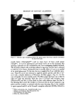
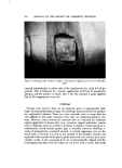
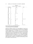
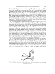
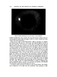
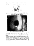
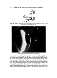
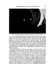
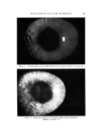
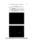
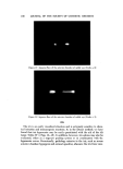
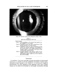
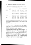
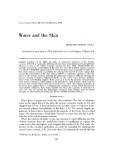
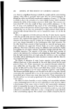
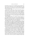
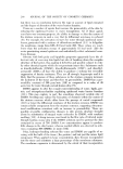
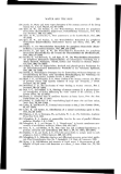
EVALUATION BY SLIT-LAMP TECHNIQUES 191 Table IV Slit-Lamp Examination of the Iris _ Iritis Grade 1 Slight congestion of iris vessels usually only part of iris involved Grade 9. Mild congestion of iris vessels, all vessels of the iris involved Grade 3 All vessels of the iris moderately congested Grade 4 All vessels of the iris congested and dilated, with very little normal tissue observable d ...½" i•-" . '2' Figure 13. Normal iris of rabbit eye (Grade = 0) The lens represents a critical structure for evaluation since any drug-related pathological change in the lens represents a substantial toxic phenomenon. Lens pathology also was not covered in the original Draize method, but is usually observed as either opacities of the anterior or posterior capsule, cor- tex, and/or some combination of all (Fig. 16). Separate grading scales may be established for each type of observed opacity. As an example of a grading system for lens opacities, Table V shows changes in anterior capsule integrity. The experience in this laboratory has been that the investigator should not integrate numerical values for each parameter into an average score. The data are best represented by calculating the mean scores for each parameter sepa- rately and indicating the incidence. An overall average score as one can cal- culate by the Draize method does not allow the investigator to illustrate or tabulate what ocular pathological change is more prominent. We no longer calculate average scores but present the data as in Table VI.
Purchased for the exclusive use of nofirst nolast (unknown)
From: SCC Media Library & Resource Center (library.scconline.org)