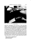
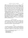
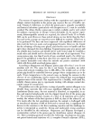
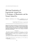
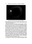
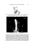
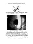
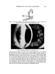
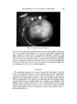
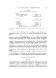
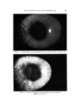
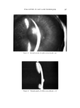
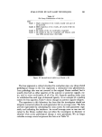
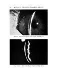
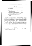
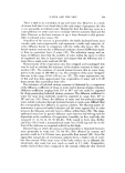
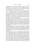
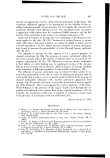
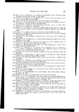
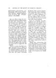
192 JOURNAL OF THE SOCIETY OF COSMETIC CHEMISTS ß. --...•' • •:,•:." •::i:iY •i-? ?. :. •...:. :.:,.:..:. i•ii: ' ß .:.•:..:... - ß ' 't:':•:' ..:.•x. .2. . .. •...}:•:•.:•:::•.. •:..... :•:•::. .... •.:.:. •:.. .:•r .•.• . ß ...... :: •.. • •,.: ::.•?' .'• . .. ... ':".... -: -• •fi• • ..... ..: ...g:.:.: :.•:• Figure 14. Iritis (congestion of iridal vessels) of rabbit eye (Grade • 2) Figure 15. Iritis of rabbit eye (Grade -----4). (Note: Hyperfolding-edema)

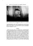
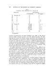
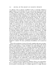
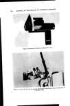
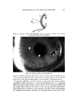
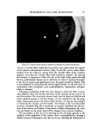
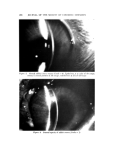
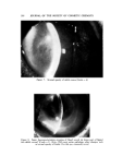
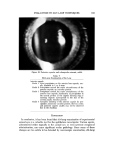
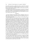
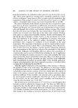
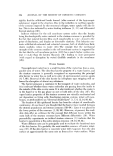
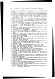
EVALUATION BY SLIT-LAMP TECHNIQUES 193 Figure 16. Posterior capsular and subcapsular cataract, rabbit Table V Slit-Lamp Examination of the Lens Anterior capsule Grade I Faint precipitates on the anterior lens capsule, usu- ally localized in I or 2 areas Grade 2 Precipitates around the entire circumference of the anterior capsule no vacuoles present Grade 3 Precipitates around the entire circumference of the anterior lens capsule localization of precipitates in the central portion of the capsule forming ring or semiring deposits (corresponds to the size of the normal pupil) vacuoles present Grade 4 Complete clouding of the anterior capsule by pre- cipitates numerous vacuoles present the lens cortex and posterior capsule usually may not be observed due to the clouding CONCLUSION In conclusion, it has been found that slit-lamp examination of experimental animal eyes is a valuable tool for the ophthalmic investigator. Various agent. s, administered either topically to the animal eye, or even systemic compound administration, can cause significant ocular pathology. Since many of these changes are too subtle to be detected by macroscopic examination, slit-lamp
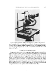
Purchased for the exclusive use of nofirst nolast (unknown)
From: SCC Media Library & Resource Center (library.scconline.org)