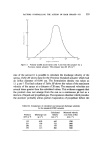
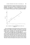
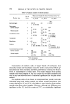
TLC STUDY OF AN ANTIMICROBIAL PRESERVATIVE 537 Separating chambers: For 20 x 20 cm plates (Desaga). UV viewer for transmission: Blak-Ray Transilluminator (Shahdon Southern Instruments Ltd). UV viewer for reflectance: Blak-Ray Chromato-Vue (Shahdon Southern Instruments Ltd). Chromoscan densitometer with thin layer attachment: Joyce Loebl and Co. Ltd. Reagents Germall 115 (Imidazolidinyl urea). Flow solvent: chloroform: methanol: acetic acid: water 50: 30: 10: 10. Spray reagent: Ninhydrin--0.3 g ninhydrin in 95 ml n-butanol and 5 ml acetic acid. Procedure Sample application 5 •tl of a 10•o solution or suspension of the sample in methanol-water (70: 30) is applied using a Drummond Microcap pipette. When the samples examined are of a viscous nature, it is necessary to use the rubber bulb in order to fill and dispel the solutions. The size of the applied spot is normally 0.5 cm diameter. Sample and reference solutions are spotted alternately along the plate at a height of 2 cm from the edge of the plate. The spots are well dried after application using a warm-air dryer. Reference solutions Solutions are prepared of the product under test without imidazolidinyl urea and the product containing varying known quantities of imidazoli- dinyl urea from 0.1 •o to 0.6•o. Chamber A Desaga chamber for 20 x 20 cm plates is lined with filter paper, satur- ated in the flow solvent and allowed to equilibrate for 30 min before use. Development The chromatogram is allowed to develop for 50 rain, in which time the solvent front travels approximately 9 cm.

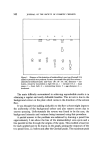
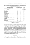
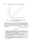
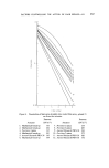
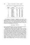
538 JOURNAL OF THE SOCIETY OF COSMETIC CHEMISTS Visualization The plate is briefly dried using a warm-air dryer and sprayed with the ninhydrin spray until well wetted. The plate is heated at a temperature of 150øC for 20 rain, allowed to cool and then viewed in uv light of 366 nm by transmission. The imidazolidinyl urea preservative is seen as two pale yellow fluorescent zones at Rfs of 0.27 and 0.35. Densitometric measurements These are performed on a Chromoscan densitometer with a thin layer attachment. The following operating conditions are used. Chromoscan: light source 12 V, 100 W, tungsten halide lamp, filter 3.00.D., optical wedge 0-0.50.D., gain 5, cam A. Thin layer attachment Light source uv mercury lamp type ST 75, aperture 1 mm x 17 mm, uv filter 300-400 nm between the light source and specimen, Kodak Wratten uv filter No. 2E between the specimen and detector, specimen expansion ratio 1: 1. The measurements are carried out using the reflectance mode. RESULTS AND DISCUSSION During the optimization of the method, various alternative systems were attempted. Laboratory-prepared plates were compared with precoated plates. The laboratory coated plates were prepared by mixing 52 g of Merck, Silica gel 'G' F254 with 110 ml of distilled water in a Waring blender and ten glass plates were then coated using a Camag automatic spreader set at a wet layer thickness of 300 •tm. The plates were left at room temperature for 20 min and finally dried for 1 h at 105øC in an air-blown oven. It was found that although satisfactory for samples containing 0.5•o imidazolidinyl urea, the laboratory-prepared plates did not display the necessary sensitivity at the lower concentrations of preservative. This may in part be due to the increased initial size of the applied spot on laboratory-prepared plates, which more readily absorb the viscous sample solutions than do the poly- vinyl alcohol bound precoated layers. Alumina laboratory-prepared plates were also examined but these failed to produce a satisfactory separation. On investigating various sample solutions, water alone usually yielded an excessive quantity of bubbles and a solution which was too viscous to pipette conveniently. The urea compound was found to be insufficiently soluble in methanol alone to be certain of recovery from emulsified products,
Purchased for the exclusive use of nofirst nolast (unknown)
From: SCC Media Library & Resource Center (library.scconline.org)