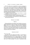
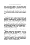
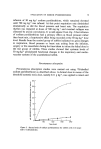
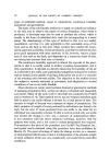
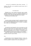
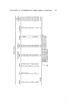
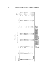
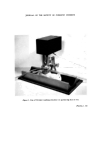
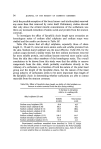
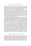
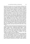
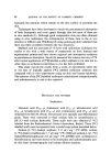
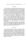
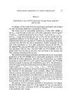
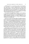
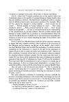
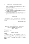
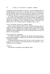
EVALUATION OF SODIUM PYRIDINETHIONE received artificial respiration whereas the other groups breathed spontan- eously for the duration of the infusion. A control group of rabbits anaes- thetized, but breathing spontaneously, was infused with a 1.8•o w/v saline solution (equivalent in tonicity to 4.0•o w/v sodium pyridinethione) at a rate of 0.5 ml rain -• for a period of time in excess of the time required to reach the lethal dose for any rabbit in either of the test groups. This experi- mental arrangement permitted the respiratory and cardiovascular systems to be monitored for the detection of functional changes at threshold dose levols. Dermal application studies All derreal applications were carried out on female New Zealand white rabbits weighing 1.6-2.5 kg. The rabbits were restrained in stocks to isolate the application site in the dorsal-lateral lumbar region. The application site was shaved 24 h prior to derreal administration and the aqueous topical application confined to an area of skin measuring 7.9 cm 2 using a moulded perspex occlusive device (Fig. 1). This device was secured to the skin by means of Stomaseal adhesive discs (Medical Products Division, 3M Com- pany, St Paul, Minnesota) and Ostomy adhesive solution (Salt & Son Ltd, Birmingham). The entire device was harnessed to the animal by an elasticated sleeve positioned around the animal's trunk. An application volume of 4.2 ml was introduced into the device through the aperture designed to accommodate a size 12 hypodermic needle. Abrasion of the application site was produced using a stripping tech- nique employing cellophane tape (6). The net effect was to produce an application site which was erythematous without showing signs of capillary bleeding. The dermal dose of asS-labelled sodium pyridinethione was 0.11 g kg -x (specific activity 0.4 gCi mg -•) in a constant volume of 4.2 ml. The topical application was left in contact with the skin for 4, 8, 12, 16, 20 and 24 h experiments, during which time blood samples were taken at 30 min inter- vals from the marginal ear vein for quantitative studies of drug serum levels. The animals were killed by cervical dislocation and exsanguinated. Per- cutaneous absorption of asS-labelled sodium pyridinethione from the site of application was quantified by a disappearance technique based on the method used by Parekh and co-workers (7). Tissues were removed, blotted and weighed in preparation for radiometric analysis.

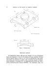
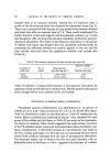
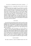
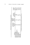
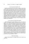
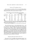
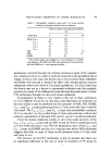
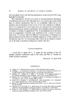
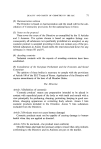
JOURNAL OF THE SOCIETY OF COSMETIC CHEMISTS Plan /'//'/ •• '• •e,• / Inlet for top•cal solution • Slots for elast•cated sleeve Side view ..... .52 mm • Figure 1. Occlusive device. Radiometric methods For distribution studies, 500 mg tissue samples or 0.5 ml of biological fluids were digested in 3 ml of 0.5 M solution of tissue solubilizer Protosol (N.E.N. Boston, Mass.) at 50 ø for 18 h. The ass radioactivity in the solubi- lized media was measured in plastic vials containing 14.5 ml of toluene- based scintillator containing 0.8• 2,5-diphenyloxazole (PPO) and 0.01•o p-bis-(2,5-phenyloxazole) benzene (POPOP). The radiometric assay was
Purchased for the exclusive use of nofirst nolast (unknown)
From: SCC Media Library & Resource Center (library.scconline.org)