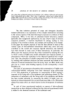
50 JOURNAL OF THE SOCIETY OF COSMETIC CHEMISTS In vitro penetration through rat skin Female Colworth-Wistar rats (100-120 g) were clipped to expose dorsal skin 24 h before cervical dislocation. The skins were excised and mounted in 2.5 cm diameter penetration cells similar to those described by Ainsworth (9). 0.25 ml of' the [•4C] surfactant solution was pipetted onto the epidermal surface of the skin and 10.0 ml of saline was added to the sampling com- partment against the dermis. The cells were kept in a warm room at 37 ø throughout the experiment and the saline was magnetically stirred con- tinuously. The saline was monitored hourly for x4C by removing 1.0 ml and replacing with fresh saline maintaining the volume of 10.0 ml in the sampling compartment. After 24 h the epidermal surface was washed with excess of distilled water and was monitored for •C by solubilizing 1 cm diameter autopsies in 'Soluene' (Packard Instruments Ltd) and counting as recom- mended by the manufacturers. In vitro penetration through human epidermis Female abdominal skin samples obtained at autopsy were frozen and stored at- 70 ø. Samples of the skin were allowed to thaw out and were heated at 58 ø for 2 min and the epidermis removed in sheets. The epidermal samples were mounted in 1 cm diameter penetration cells similar to those described by Ainsworth (9). Saline containing 0.012•o Pencillin and 0.01• Streptomycin was placed in contact with both surfaces of the sample and the cells were equilibrated at 37 ø for 24 h. The electrical resistance of the cells was measured and only cells with a resistance greater than 50 000 12 were used. The saline from the corneum surface was removed and 0.1 ml of the [•4C] surfactant solution was placed on the corneum. 1.0 ml aliquots of the saline in the sampling compartment (8.0 ml) were monitored for •C at 0.5, 1, 2, 3, 4, 6, 7, 8, 24 and 48 h, each time 1.0ml of fresh saline was added to maintain the volume at 8.0 ml. At the end of the experiment the comeurn was washed with excess of distilled water and the epidermal sample monitored for x•C by solubilizing in 'Soluene'. Animals and treatment Female Colworth-Wistar rats weighing 100-120 g were used for all experiments.

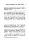
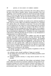
PERCUTANEOUS ABSORPTION OF ANION1C SURFACTANTS 51 Turnover of surfactants The turnover of each [x4C] labelled surfactant was measured by injecting three animals intraperitoneally and three animals subcutaneously with 0.1 or 0.5 ml of surfactant solution. The animals were then placed in sealed metabolism cages where urine, faeces and expired air were collected and monitored for x4C. The metabolism cages consisted of airtight perspex cages mounted on polythene collection funnels which directed the excreta into 'Metabowl' urine/faeces separators (Jencons Ltd, Hemel Hempstead, Herts). Air was drawn through the cages at 1.5 1 min 4 and bubbled through towers 30 cm deep and containing 240 ml of 50•o aqueous ethanolamine. 1.0 ml aliquots of this solution were monitored for •4C at regular time intervals. Each urinary sample was made up to 25 ml with cage rinsings and faecal samples were freeze-dried. After 6 or 24 h the animals were killed by cervical dislocation. The carcasses of the animals were homogenized in an 'Atomix' blender (M.S.E. Ltd, Crawley, Sussex) and aliquots of the homogenate were freeze dried. Percutaneous absorption The hair from animals' backs was removed with fine bladed clippers 24 h before topical application. Only animals with visibly undamaged skin were used in the topical studies and all animals were lightly anaesthetized with a cyclopropane: carbon dioxide: oxygen gas mixture during treatment. Topical application of 0.1 or 0.5 ml of the [•C] test solution was made from a microlitre syringe on to an area of skin (7.5 or 10 cm 2) previously marked out on the animal's back with a felt-tipped pen. The solution was lathered over the treatment area with a rounded glass rod for 1 min during application. After 15 min contact with the skin the animal was inverted over a 6-inch diameter funnel and the excess of test solution was rinsed off with distilled water at 37 ø from a wash bottle. After about 50 ml of water had been used the treated area of skin was lightly drawn over the top of the funnel to squeeze excess of rinse water from the skin. This process was then repeated and the skin dried with paper tissues. The animals were then fitted with either restraining collars or non-occlusive protection patches and placed in the metabolism cages for collection of excreta as described above.
Purchased for the exclusive use of nofirst nolast (unknown)
From: SCC Media Library & Resource Center (library.scconline.org)