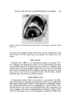
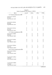
]. Soc. Cosmet. Chem., 26, 411-425 (August 1975) An In Vivo Method for the Detection of Residual Antimicrobial Activity on Human Skin EDWARD EIGEN, M.A., ALADAR LEGENYEI, and SIDNEY WEISS, Ph.D.* Presented May 10, 1974, SCC Seminar, Chicago, Illinois Synopsis-A new IN VIVO METHOD has been devised, which realistically estimates RESIDUAL ANTIMICRO•BIAL ACTIVITY on skin. Residual activity is measured by placing a small petri-type dish containing a DIFFERENTIAL MEDIUM seeded with a known amount of one specific organism on a treated site for 4 hours. The dish is then incubated for 48 hours, and the colonies, which represent survivors, are counted. These counts are compared with counts obtained in the same manner from an area treated with a PLACEBO. The duration of antimicrobial activity on the skin is followed in this man- ner over a period of several days. Using this technique, it has been possible to demonstrate differences in residual activity remaining on the skin after application of liquid antimicro- bial skin cleansers and antimicrobial soap followed by thorough rinsing with water. INTRODUCTION Some methods for determining residual antimicrobial activity on skin de- pend on microbial counts before and after treatment, or extraction of skin with solvents and analysis of the extracts for the presence of the antimicrobial agent. The number of techniques available shows how difficult this task is. Typical procedures dependent on bacterial counts include: swabbing (1) contact plate ( 1) tape-stripping (2) and washing in basins (3, 4). Each has error because not all the organisms on a test site can be collected or counted. Other complications include the type and number of organisms indigenous to *Colgate-Palmolive Research Center, Piscataway, N.J. 411

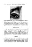
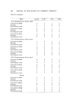
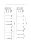
412 JOURNAL OF THE SOCIETY OF COSMETIC CHEMISTS the treated sites and individual differences among subjects. In addition to these problems, Shaw et al. (5) found additional variables influencing the counts to be day-to-day variation in bacterial populations at the same site, differences in population in adjacent areas, and the methods used to collect bacteria. The use of only one medium to count bacteria yields partial informa- tion since it excludes organisms which will not grow in that medium or at the temperature selected for incubation. Other methods for measuring the amount of agent deposited on viable skin have been reviewed by Taber et al. (6). These include solvent extraction followed by either spectrophotometric or microbiological assay, tape-stripping and examination for particles on the tape, and use of radioactive materials. These methods can also be subject to error in two ways. First, extraction may not be complete and second, measurement for the presence of a compound does not indicate whether the material retained its antimicrobial activity when adsorbed on the skin. In the case of surface counting of radioactive compounds, counting efficiency has been reported by Taber et al. (6) to be 4.5% for •4C. This suggests that low concentrations of •4C tagged compounds cannot be measured in situ unless the specific activity is high. Tritiated com- pounds might be almost impossible to study since the efficiency of counting is much lower. In addition, radioactive compounds are not always available. A method to determine residual activity on the skin, in vivo, that avoids many of these variables is highly desirable. EXPERI1ViENTAL In considering this problem, it is logical to assume that treatment of the skin with a product capable of killing or inhibiting the growth of certain organisms does in fact accomplish this task. What is being sought, therefore, is evidence that this effect lasts for some period of time so that if all the organisms on these same areas are not killed, or if the site is invaded by the same type of bacteria, they will not be able to grow. It is also of interest to obtain some quantitative estimate as to the efficacy of the residue on the skin. A method has been devised which yields a quantifiable estimate of residual antimicrobial activity on the skin. The method consists of the following steps. 1. A particular organism is grown in a suitable liquid medium. 2. The culture is standardized and diluted with normal saline to contain an appropriate number of organisms per milliliter. 3. Small seeded agar plates are prepared which contain the desired num- ber of organisms per plate. 4. These plates are placed on discrete areas of the body which have been pretreated with an antimicrobial preparation. Care is taken to make sure that the entire agar surface is in contact with the skin. The plates are secured and allowed to remain in place for 4 hours.
Purchased for the exclusive use of nofirst nolast (unknown)
From: SCC Media Library & Resource Center (library.scconline.org)