THE NATURE OF DANDRUFF 125 in the practice of dividing the scalp into a number of segments, with each one receiving a separate grade. One scheme calls for separate evaluations of 25 areas, a 20 to 30 min process (4)! Some delineate 9 areas while the "short" method utilizes 4. We compared the corneocyte counts on opposite sides of the scalp in 140 individuals with and without dandruff. The correlation between the two sides was exceptionally high (r--0.843). A phenomenally straight line was pro- duced when the right and left sides were plotted against each other. There was no evidence of asymmetry. Also, by clinical grading, dandruff seems to us to be a uniformly diffused (not patchy) process. Seborrheic dermatitis, on the other hand, presents itself in the form of circumscribed lesions with indistinct borders, just as it does on the skin. It, not dandruff, is patchy. Microorganisms recognize the difference between dandruff and seborrheic dermatitis. While small numbers of S. aureus occassionally can be recovered from dandruff, this potentially virulent organism can be isolated in large num- bers from 20 per cent of the subjects with seborrheic dermatitis (9). S. aureus may comprise as much as one-third of the total flora. Finally, dandruff wanes with age, while seborrheic dermatitis increases. X. HISTOPATHOLOGY No aspect of our study has taken more twists and turns than the views we have held at different times concerning the microscopic changes in dandruff. It will be instructive to review the origins of this confusion. We were satisfied after studying about 50 dandruff specimens that we had a good grasp of the pathologic changes (10). We had examined many normal scalps in the past, and thought we knew the terrain, but our attention then was on the hair follicle, not the epidermis. Naturally, it is quite easy not to see when one is not looking. For a while, we thought that there was in dandruff an increased perivascular infiltrate of mononuclear cells in the upper dermis, a kind of smouldering low grade chronic i•ffiammatory reaction. The idea ap- pealed to us, since at that time, we believed that dandruff was merely mild seborrheic dermatitis. Experts in cutaneous histopathology, who looked at these specimens, agreed with our interpretation, but, of course, they too had no real experience with the anatomy of normal scalps. It is not until one has studied scores of so-called normal scalps that one begins to appreciate the extraordinary individual variations. Many of these show round cells infiltrates to a similar degree. We came to realize that the cuff of adventitial cells, which normally surround small vessels, was greater over the head. The microvascu- lature of the scalp is very richly developed, and mononuclear cells seem to emigrate into the tissues more readily, a sort of slow extravascular circulation. We made the same mistake in studying ache of the face, where, again, nor- mally there are more mononuclear cells patrolling the tissue, creating a spe- cious appearance of low-grade i•ffiammation.

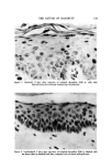
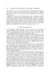
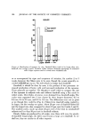
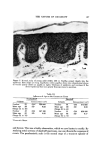
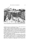
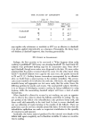
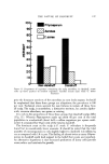
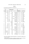
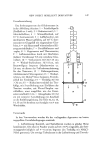
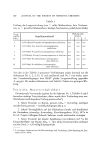
126 JOUBNAL OF THE SOCIETY OF COSMETIC CHEMISTS On the scalp, the undersurface of the normal epidermis is very strongly sculptured. The dermal papillae deeply dent the epidermis so that in cross- section the dermo-epidermal contour is highly undulating with long rete pegs (Fig. 5). Sometimes the appearance is psoriasiform. Initially, we thought that the epidermis in dandruff was thicker (acanthotic). This, too, turned out to be a variable feature and not at all characteristic of dandruff. An important fac- tor in this regard is age. Deeply projecting papillae with long rete pegs are very characteristic of youth. After middle age, the dermo-epidermal contour progressively flattens and the thickness of the epidermis decreases. In old age, the dermo-epidermal junction is flat rather than wavy, and the epidermis may become very thin. These changes, along with decreased cell turnover, explain the disappearance of dandruff in the aged. It is appropriate, here, to provide objective data on the extent to which age influences the rate of production of horny cells. Using corneocyte counts, we compared elderly subjects with and without dandruff to young adults. By cli- nical criteria, dandruff was equally severe in both groups (Grade V). It turned out, however, that the counts were appreciably lower for the elderly (Table III). Within each age group, the dandruff subjects had higher counts, the difference being especially prominent in the aged. This is another illustration of lack of a strict correlation between counts and grades. Apparently, the elderly can produce just as many scales even though cell turnover is de- creased. Since scaling is the central feature of dandruff, one might think this would be a dominant finding in histologic sections. Unfortunately, fixation and pro- cessing literally ruins the horny layer. Judgments of thickness and quality of the SC are very hazardous. Quite often, it is simply knocked off in sectioning -leaving only fragments. Originally, we thought that there was a diagnostic histologic feature in dandruff. We said, "the hallmark of dandruff is scattered loci of parakeratosis" (10) (parakeratotic horny cells retain their nuclei). Small mounds of paraker- atotic horny cells are to be sureco mmon in dandruff, but identical findings are present in nondandruff. Again, the difference is merely quantitative. To find them in nondandruff may require examination of many sections. They be- come increasingly sparse as the grades diminish from III to I. The reader has no doubt begun to sense that the parakeratotic loci are, in fact, the visible flakes or squames, which are universal in all scalps. Like the corneocyte count, their quantity is proportional to the clinical grade. Paraker- atosis invariably signals increased prolferative activity and is usually a con- sequence of underlying inflammatory change. Parakeratosis is typical of such inflammatory dermatoses such as seborrheic dermatitis and psoriasis in which cell turnover is sharply accelerated. In our previous work, we were mystified as to how these parakeratotic mounds formed in dandruff. They often seemed to be sitting over a normal epidermis
Purchased for the exclusive use of nofirst nolast (unknown)
From: SCC Media Library & Resource Center (library.scconline.org)