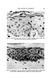
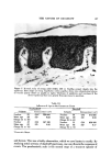
THE NATURE OF DANDRUFF 117 out of the basal zone and in their upward passage undergo a complex series of transformations designated by the general term, 'differentiation' and by the dermatologic term, 'keratinization.' As each cell becomes larger, tonofila- ments (bundles of the fibrous protein keratin) increase in quantity and thick- ness, new organelles appear, the cell membranes thicken, and the eelIs be- come increasingly stuck to one another. All these and many other changes, which take place in the differentiating compartment, have a specific biologic goal, the production of horny cells. The latter comprise the fabric of the stratum corneum (SC). Horny cells or corneocytes are dead rigid cells, literally bags of fibrous protein encased in membranes so tough that strong alkalis dissolve out the cellular contents leaving empty sacs. The horny cells are held together by a strong intercellular glue, forming thereby a coherent horny layer The func- tion of the latter is to act as a "barrier" to prevent the passage of substances into or out of the skin. The SC seals off the body from the environment. Near the surface, the SC begins to crack. This is the desquamating zone, where the cells be- come loosened in preparation for their being shed. This outer loose porous zone is only three to four eel] layers thick. It is important to understand that horny cells do not come off individually, but in variably sized aggregates, comprising tens and even hundreds of cells. These clumps are, for the most part, invisible, being less than 200/xm (0.2 mm) in diameter. The individual horny cells are about 40 /xm in diameter. Aggregates larger than about 0.2 mm are visible to the naked eye as flakes or squames. Depending on the thickness, flakes as large as 2 mm may contain thousands of horny cells. Very large flakes may contain hundreds of thousands of cells. Squames are, of course, the hallmark of dandruff they become more numerous and larger with increasing severity. We shall have to be concerned with their origin. One of the first questions which arises is whether dandruff subjects pro- duce a greater quantity of horny cells. Or are corneocytes merely being shed in visible flakes? Some measurement of epidermal proliferative activity is re- quired. Specialists concerned with epidermopoesis have developed various techniques for estimating the rate at which the epidermis renews itself. One can determine the average time for a cell to move from the basal layer to the surface (the transit time). Indeed, it is possible to calculate separately re- newal times for the dead horny layer and the viable epidermis. None of these measurements have been made for the scalp. Still, we have generated some data which enables a meaningful comparison of epidermal kinetics in persons with and without dandruff. In the studies summarized below, the subjects were always young adult males. The mitotic index, the percentage of basal cells in mitosis, affords an esti- mate of how rapidly the celts in the germinative conapartment are reproduc-

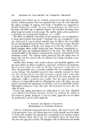
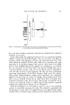
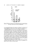
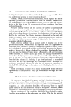
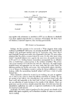
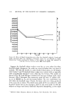
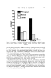
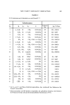
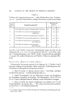
118 JOURNAL OF THE SOCIETY OF COSMETIC CHEMISTS Table I Mitoses Per Thousand Basal Cells Subjects Nondandruff Grades II and III Subjects Dandruff Grades V and VI 14 Mean 14.6 18 Mean 30.6 SD '• ___4.9 SD • ___7.9 :'Standard deviation. Table II Labeling Index (Number of Labeled Cells l•er 100 Basal Cells) S •bjects Nondandruff Grades II and III Subjects Dandruff Grades V and VI l0 Mean 11.0 l0 Mean 17.5 SD -t-2.4 SD ___4.4 ing themselves. Because it is difficult to identify cells in early and late mito- sis, this determination is rather imprecise. Since only a small percentage of basal cells are dividing at a given time, one must scan thousands of cells to keep the experimental error within bounds. Comparisons were made between 14 nondandruff subjects (Grade II and III) and 15 dandruff subjects (14 Grade V and 4 Grade VI). The results are shown in Table I. Even though the values were distributed over a wide range, the mitotic index was about twice as great in the dandruff group (p = 0.01). This result indicates that proliferative activity is increased in dan- druff. This was the first evidence we secured that more cells were being shed from dandruff scalps. It is worth pointing out here that cell turnover on the head region is normally much swifter than on the glabrous skin (6). Another means of estimating cell turnover is to "tag" cells in the reproduc- tive compartment with tritiated thymidine. Those cells which are in the DNA- systhesis phase of the cell cycle will incorporate the radioactive nucleotide into their nuclei. After suitable histologic processing, one can determine the percentage of radio-labeled basal cells (the labeling index). The technique entails a 0.1-ml intradermal injection of 5 to 10 microcuries of tritiated thymi- dine followed by excision biopsy 45 min later. The number of labeled cells per hundred basal cells was determined in 16 subjects without dandruff (Grades II and III) and 19 subjects with Grades V and VI. The results are shown in Table II. Again, the range was great within each group. There can be little doubt, however, that the proportion of DNA-synthesizing cells was about twice as large in dandruff, indicating much faster cell renewal. The rate at which labeled cells move through the epidermis in their journey to the surface (transit time) is another indicator of proliferative activity. To follow cell movement, one delays excising the injected area for a variable number of days. One can then see how far up the tagged cells have migrated
Purchased for the exclusive use of nofirst nolast (unknown)
From: SCC Media Library & Resource Center (library.scconline.org)