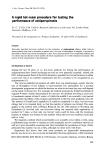
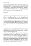
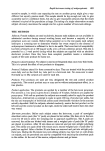
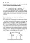
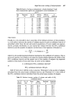
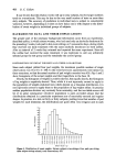
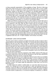
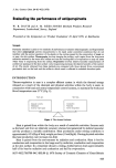
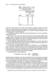
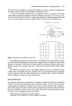
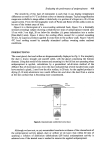
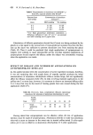
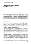
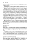
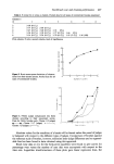
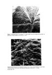
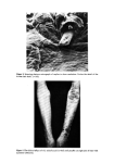
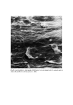
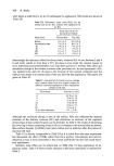
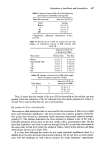
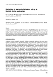
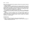
Figure 1. Scanning electron micrograph of a replica taken from skin treated with an HA 2 h previously. Notice shallow skin furrows. (x 16.5). v.. .- .:. •,, . • , .... . , • . ..... • • • •-C ' ..... '-• -.. .. ..... .- • • • . -•-•. " .•.. •'•.' -•..%. •'•'. •... 2 • • •,: •.• •.'.• •,• .• -,•--: -.-•,• . ...... • .i.... ".•'• • - ...... . . - , - .•. .:% •-•. : • . . • . • •.' - '•'•,•.?' %.... •? ...? - . ..... . •.-..:. Figure •. Scanning electron micro•aph of replica of skin surface treated with HA 2 h previously. Notice prominent cell borders and thickened individual corneocytes. (x 1-28 K). Facing page 43•

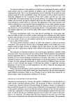
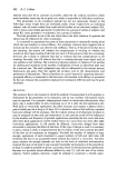
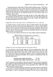
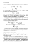
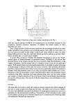
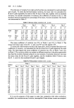
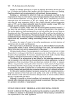
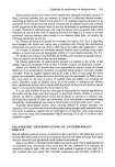
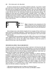
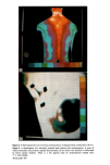
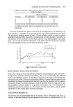
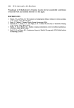
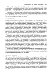
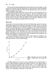
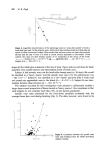
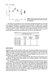
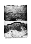
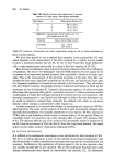
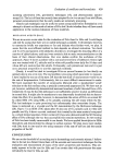
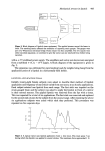
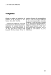
Figure 3. Scanning electron micrograph of replica to show resolution. Notice the detail of the broken hair shaft. (x 165). Figure 4. The clinical effects of 6% salicylic acid in white soft paraffin on right arm of man with Lamellar ichthyosis.
Purchased for the exclusive use of nofirst nolast (unknown)
From: SCC Media Library & Resource Center (library.scconline.org)