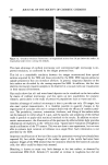
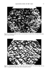
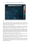
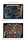
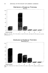
j. Soc. Cosmet. Chem., 44, 1-12 (January/February 1993) 3D Reconstruction of human hair by confocal microscopy PIERRE CORCUFF, PHILIPPE GREMILLET, MICHEL JOURLIN, YOLANDA DUVAULT, FREDERIC LEROY, and JEAN-LUC LEVEQUE, L'Orgal, Laboratoires de Recherche Fona•mentale, Aulnay-sous-Bois, France (P.C., Y.D., F.L.,J.L.L.), Misis Image, Saint-Etienne, France (P.G.), and Letia-ICPI, Lyon, France (M.J.). Received August 31, 1992. Synopsis Confocal microscopy is a highly original, non-destructive method for observing objects in three dimensions and in their natural environment. As such, it is well suited to studies of human hair. The tandem-scanning reflected-light microscope was used to study the hair surface in its natural environ- ment, which included sweat and sebum. Micrometric measurements assessed the effects of classical treat- ments (permanent waving and bleaching), swelling in water and urea solution, and stretching. A confocal laser scanning microscope was used to reconstruct a volume of hair shaft. The internal structures (cortex and medulla) were contrasted using a fluorescent marker (rhodamin) and could be observed in both longitudinal and transverse optical sections at all levels. Our results demonstrate the value of confocal microscopy in describing hair structure and the effects of treatments on the cuticle. Methods based on classical optical and electron microscopy are far more complex and time-consuming than confocal microscopy, which represents an extremely promising technique for cosmetic research. INTRODUCTION The cylindrical shape of hair precludes straightforward observation of its surface by means of light microscopy, particularly at high magnifications. High-resolution images are therefore generally obtained by scanning electron microscopy (SEM), but the high vacuum and gold coating required prevent actual visualization of the cuticle (1). Internal structures (cortex and medulla) are classically studied on serial sections, which, needless to say, involve destruction of the sample. Confocal microscopy (2) provides high- resolution images with virtually no out-of-focus areas. In addition, the optical section- ing property preserves the integrity of the sample, even though it is limited by the opacity of the specimen, light-absorbing structures, surface reflections, refraction/ scattering, and the design of the objective lens (numerical aperture and working dis- tance). A series of optical sections is acquired and stored for later 3D reconstruction or computation of volume.

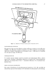
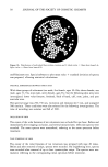
2 JOURNAL OF THE SOCIETY OF COSMETIC CHEMISTS In this study, we used two confocal microscopes to study the surface aspect and internal structure of human hair. The tandem-scanning reflected-light microscope (TSM), in- vented by Petran eta/. (3), gave high-resolution images of the surface, and was used to view and quantify the effects of cosmetic treatments, swelling and stretching on the topography of cuticular cells. Internal structures were visualized using a confocal laser scanning microscope (CLSM). Fine details of the cortex and medulla were identified by the use of a fluorochrome. MATERIALS AND METHODS HAIR SAMPLING Freshly plucked hair and eyelash were simply collected on the authors at the time of the experiment. In all other cases, commercially available Caucasian virgin brown hair was used in standardized conditions. HAIR TREATMENTS (4) A permanent wave treatment was comprised of two steps: reduction and oxidation. Reduction was done with ammonium thioglycolate (1 M), adjusted at pH 9 with ammonia, for 30 minutes at 30øC. Hair was rinsed and fixed with a 2.5% hydrogen peroxide solution adjusted at pH 3 with hydrochloric acid. Finally, samples were rinsed with tap water. Bleaching was performed by immersing virgin hair in hydrogen peroxide (6%)/sodium persulfate (10%) solution, adjusted to pH 10 with ammonia for 45 minutes at 35øC, and then thoroughly rinsing with tap water. Swelling was obtained by immersing virgin hair in either deionized water or 8 M urea at room temperature for ten minutes. STAINING WITH FLUOROCHROMES 10 mg of virgin hair were immersed for one hour in 5 ml of 0.05% solutions of rhodamin B in water or octadecyl-rhodamin in ethanol/water (9:1) at 60øC. The excess of fluorochrome was removed by dipping hairs for ten seconds in tetrachloroethylene the hair was then dried in an oven at 105øC overnight. BASIC PRINCIPLE OF THE CONFOCAL MICROSCOPE Whereas the classic light microscope homogeneously illuminates a large area within the sample, the confocal microscope acts with a focused beam of light. The reflected light at the focused point does go through a pinhole in front of a detector (TV camera, photo multiplier). Out-of-focus reflections cannot pass the pinhole and consequently are not imaged. An image is generated at the focal plane by scanning the light spot in X and Y directions with either rotating mirrors or a Nipkow disc. The image does not contain any information from above and beneath the focal plane and is called an optical section.
Purchased for the exclusive use of nofirst nolast (unknown)
From: SCC Media Library & Resource Center (library.scconline.org)