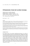
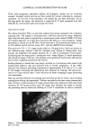
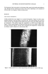
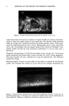
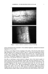
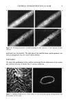
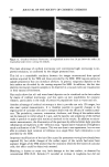
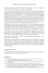
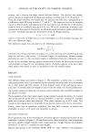
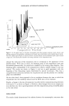
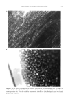
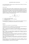
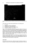
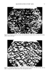
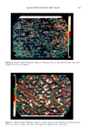
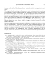
CONFOCAL 3D RECONSTRUCTION OF HAIR 9 Figure 10. Stretching experiment: a) before stretching b) 10% extension c) 15% extension d) 20% extension. penetrated into the medulla. The right part of the medulla shows typical septation the cortical arrangement was even more clearly defined. DISCUSSION We tested the performance of the confocal microscope for the observation of the surface and internal structure of human hair in various conditions. Figure 11. Rhodamin B fluorescence: a) hair surface b) cross section showing that the fluorochrome did not penetrate into the cortex.

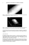
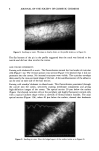
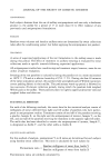
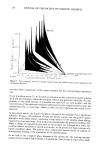
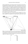
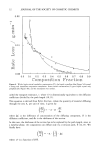
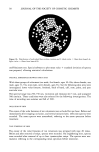
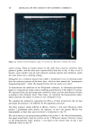
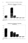
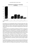
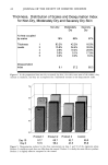
10 JOURNAL OF THE SOCIETY OF COSMETIC CHEMISTS Figure 12. Octadecyl rhodamin fluorescence: a) longitudinal section from 20 Izm below the surface b) longitudinal axial section cutting the medulla. The main advantage of confocal microscopy over conventional light microscopy is im- proved resolution, as confirmed by the images presented here. This led to a remarkable similarity between the images reconstructed from optical sections acquired by the TSM and those provided by the SEM. SEM requires extensive sample preparation that can introduce artefacts. In general, exogenous deposits on the hair surface are disturbed by the coating required for SEM. In addition, the fact that electron microscopy requires samples to be observed in a vacuum rules out visualization in their natural environment. Our results show that oil- and water-based deposits can be visualized on the hair surface by means of confocal microscopy, and this opens up new possibilities for cosmetic research, particularly in the study of protective preparations such as waxes and oils. Another advantage of confocal microscopy is that it provides not only 3D images, but also exact spatial measurements. It is therefore possible to quantify changes in the organization of cuticular cells and to compare objectively the efficacy of cosmetic prod- ucts. The periodicity, contours, orientation, thickness, and lifting of the surface scales can be measured to within about 0.1 Ixm, and the rapidity and simplicity of the method make it possible to apply valid statistical methods to the results. In addition to micro- metric measurements, the illumination of the specimen by reflected white light provides information on brightness in the "reflection map." Quantitative evaluation of brightness needs a calibration of the TV camera. This was not done in our experiment, but we were able to evaluate local variation in brilliance on a single fiber. Such information is not provided by the SEM. Important modifications of the hair fiber caused by permanent waving have already been reported (8), but subtle changes of the surface aspect of the cuticle had never been assessed. Images of the TSM showed that the treatment lifted the free border of cuticular cells this effect could be objectively assessed. Bleaching is known to cause very little damage to the hair surface, as observed by conventional SEM (8,9). Zelinski (7) recorded an increase in roughness but did not
Purchased for the exclusive use of nofirst nolast (unknown)
From: SCC Media Library & Resource Center (library.scconline.org)