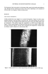
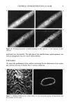
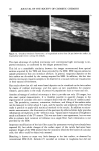
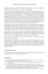
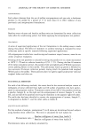
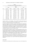
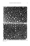
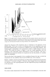
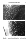
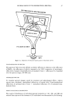
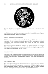
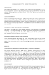
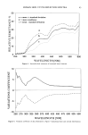
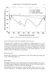
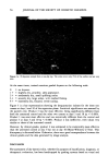
38 JOURNAL OF THE SOCIETY OF COSMETIC CHEMISTS Figure lb. Distribution of individual fibers within common end C (dark circles = fibers from branch A light circles = fibers from branch B). oroff-Smirnow test. Special software to plot mean value q- standard deviation of spectra was prepared, allowing statistical calculations. SPECTRAL DIFFERENCES BETWEEN BODY SITES With three groups of volunteers (ten male, five female, ages 10-20a eleven female, one male, ages 31-55a nine male, seven female, ages 56-72a) the following skin sites were investigated: lower volar forearm, forehead, back of hand, calf, nose, palm, and post auricular area. The spectral range was 250-750 nm, excitation and emission slit 3 nm, and scanspeed 500 nm/min. These conditions were also selected for the following investigations. The time of recording was summer and fall of 1991. OCCLUSION OF SKIN The center of the volar forearms of ten volunteers was occluded for one hour. Before and immediately after stopping occlusion, and several minutes later, reflection spectra were recorded. The mean spectra were normalized, referring to the mean spectrum before treatment. TAPE STRIPPING OF SKIN The center of the volar forearms of ten volunteers was stripped with tape 20 times. Before and after removal of strips, spectra were recorded. On neighboring sites, spectra were recorded after removal of up to four cyanoacrylate strips. The spectra were nor- malized, referring to the corresponding mean spectrum before treatment.

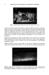
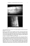
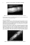
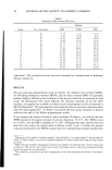
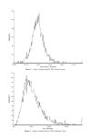
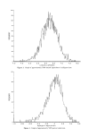
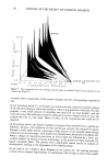
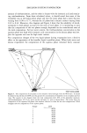
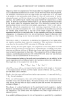
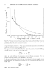
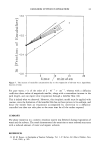
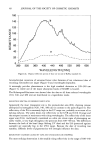
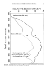
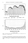
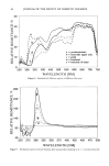
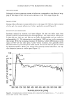
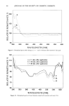
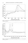
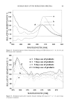
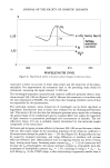
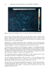
HUMAN SKIN UV/VIS REFLECTION SPECTRA 39 DRYING OF SKIN The middle volar forearm of five volunteers (three female, two male mean age = 25 + 4.2a) was dried by means of dry silica gel under occluding polyethylene film for 60 minutes. Before application and after removal of the film, spectra were recorded and normalized. CHANGING BLOODFLOW On the volar forearm of five volunteers, erythema was induced by pressure (dermograph- ism), nicotinic acid benzylester (1% in ethanol), and infrared radiation. Reflectivity was measured before and after applying pressure on the erythematous skin. Venous occlusion was induced by interrupting circulation: two and five minutes later reflection spectra were recorded. INFLUENCE OF A SINGLE TREATMENT WITH SURFACTANTS Tap water (3 ml), soap slurry (10% aqueous dispersion, 3 ml) and SDS (10% aqueous dispersion, 3 ml) were applied for ten minutes on the volar forearms of ten volunteers under a funnel pressed against the skin surface. Before and 1, 10, 20, and 40 minutes after treatment, reflection spectra were recorded. INFLUENCE OF FREQUENT WASHING WITH DETERGENTS Thirty volunteers washed their hands and lower forearms four times per day for two minutes each and for two weeks with a classic soap bar free of perfume and refattening agents. Two, seven, nine, and 14 days after starting treatment, reflection spectra were recorded. RESULTS STANDARDIZATION OF SPECTRA AND IDENTIFICATION OF ENDOGENOUS ABSORBERS The original reflection spectra of BaSO 4 and of skin look similar (Figure 2a). After standardization, the relative spectrum is comparable to formerly published spectra (6) (Figure 2b), with exception of the UV range. It is obvious that reflections are missing (=absorption) in the range of fluorescent NADH (h. max 340-350 nm) in the UVB/ C-range, where aromatic amino acids, especially tryptophan, are emitting fluorecence and around 400 nm, where endogenous and bacterial porphyrins are efficient, fluores- cent absorbers located at special skin sites (nose, upper back). Most of the observed absorption peaks can be related to definite endogenous epidermal or dermal chro- mophores (1). REPRODUCIBILITY The intraindividual variation coefficient determined from ten spectra gained within 15 minutes from the same skin site is approximately 2% from 300-650 nm, showing higher deviations especially in the deep red portion of the spectrum (Figure 3).
Purchased for the exclusive use of nofirst nolast (unknown)
From: SCC Media Library & Resource Center (library.scconline.org)