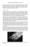
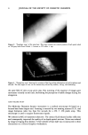
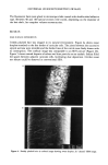
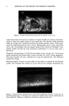
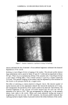
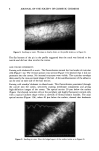
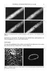
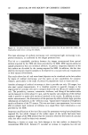
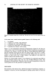
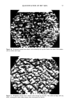
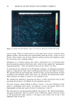
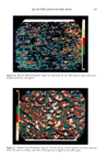
CONFOCAL 3D RECONSTRUCTION OF HAIR 11 interpret his observation. With the confocal microscope, we were able to confirm the corrosive effect of the treatment, as previously suggested (8). The simplicity of this imaging method and its non-destructive nature open the way to studies of dynamic phenomena in four dimensions. This is illustrated by our study of swelling and stretching. Although hair swelling has previously been studied by means of physical techniques, this is the first time, to our knowledge, that periodic phenomena (bulging) have been described. The same is true of the stretching studies. Although measurements of resistance to traction have classically been used to evaluate the visco- elastic properties of hair, changes in surface structure during stretching have rarely been reported because of technical difficulties inherent in conventional microscopy. The variations we observed in the brightness of the borders of cuticular cells during stretch- ing suggest their rising as previously reported by Brown and Swift (10). Hair specialists are well aware of the fastidious and time-consuming nature of methods for preparing longitudinal and transverse sections. Even the most skilled technicians rarely produce sections that are perfectly aligned with the hair axis. The optical sec- tioning property of the confocal microscope overcomes these problems, since sections can be visualized at any given level and in any given direction in a fraction of a second. The images of the internal hair structures obtained after labeling with a fluorescent marker were also of exceptional quality, compared to those obtained by observation of fiber sections in conventional fluorescence microscopy (11). Finer details of the structure of the cortical cells were observed, both in the radial and axial directions. This technique also provides a simple tool for monitoring the penetration of dyes throughout the hair thickness. In conclusion, confocal microscopy is highly suited to studies of human hair and the effects of cosmetic preparations. It is particularly adapted to observations of rounded surfaces and is thus a valuable alternative to classical microscopic methods. A particular advantage is the non-destructive nature of the technique, which means that samples can be observed both before and after treatment this opens up exciting new possibilities for cosmetic research. ACKNOWLEDGMENTS We are grateful to R. Aslund from Molecular Dynamics (Sunnyvale, CA) for providing the images obtained with the Sarastro microscope. REFERENCES (1) J. A. Swift, Fine details on the surface of human hair, lnt. J. Cosmet. Sci., 13, 143-159 (1991). (2) T. Wilson, Confocal Microscopy (Academic Press, London, 1990). (3) M. Petran, M. Hadravsky, and A. Boyde, The tandem scanning reflected light microscope, Scanning, 7, 97-108 (1985). (4) C. Zviak, The science of hair care, Dermatology, Vol. 7 (Marcel Dekker, New-York, 1986). (5) N. Aslund, K. Carlsson, A. Liljeborg, and L. MajliSf. Phoibos, a microscope scanner designed for micro-fluorometric applications, using laser induced fluorescence. Proceedings of the 3rd Scandinavian Conference on Image Analysis (Student Litteratur, Lund, Sweden, 1983), pp. 338-343. (6) C. R. Robbins, in Chemical and Physical Behavior of Human Hair (Springer-Verlag, New York, 1988).

12 JOURNAL OF THE SOCIETY OF COSMETIC CHEMISTS (7) M. Zielinski, A new approach to hair surface topography: Fourier transform and fractal analysis,J. Soc. Cosmet. Chem., 40, 173-189 (1989). (8) J. A. Swift, and A. C. Brown, The critical determination of fine changes in the surface architecture of human hair due to cosmetic treatment, J. Soc. Cosmet. Chem., 23, 695-702 (1972). (9) N. B. Fair and B. S. Gupta, Proceedings of the 7th Int. Wool Textile Research Conference, Tokyo, Vol. IV, 163-172 (1985). (t0) A. C. Brown and J. A. Swift, Hair breakage: The scanning electron microscope as a diagnostic tool, J. Soc. Cosmet. Chem., 26, 289-297 (1975). (lt) V. Sideris, L. A. Holt, and I. H. Leaver, A microscopical study of the pathway of diffusion of rhodamine B and octadecyl-rhodamine B into wool fibres,J. Soc. Dyers Colour, 106, 131-135 (1990).
Purchased for the exclusive use of nofirst nolast (unknown)
From: SCC Media Library & Resource Center (library.scconline.org)