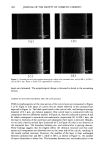
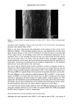
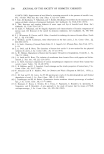
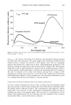
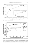
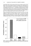
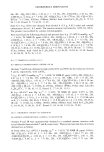
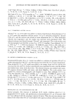
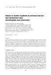
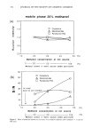
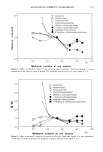
ISOLATION OF CUTICLE 283 mediately washed with ethanol containing 25% HC1, ethanol, and finally distilled water. The solution was renewed five times to remove KOH and 1-butanol completely. SCANNING ELECTRON MICROSCOPY AND TRANSMISSION ELECTRON MICROSCOPY Vertical views of the untreated and treated specimens were observed in a Hitachi S-4100 scanning electron microscope (SEM). The average diameter and the thickness of the cuticle cells from the cross-sectional views were also measured on more than thirty monofilaments of the respective specimens. TRANSMISSION ELECTRON MICROSCOPY Cross-sectional views of cuticle cells were observed by a JEOL 100SX transmission elec- tron microscope (TEM). Specimens were embedded in epoxy resin, and sections were cut on an ultra thin-cut microtome. FT-IR SPECTROSCOPY The IR spectra of the untreated and treated hairs were measured using an FT-IR microscope (Shimazu, IMS-800, Japan) and an FT-IR spectrometer in diffuse reflectance measurements (Shimazu, FTIR-8100M, Japan). To get the thin, flat section of exposed cortical cells, the monofilament was cut longitudinally on both sides. A transmission spectrum of 15 •m X 15 •m/square area of cortex was obtained with the FT-IR microscope (200 scans at 4 cm- • resolution) and an MCT detector. The spectra of the surface of the hair were obtained by scratching the surface of hair carefully with a blade. The scraped section was collected and diffuse reflectance measurements were carried out. The spectra analyzed were 200 scans at 4 cm- • resolution. In diffuse reflectance measurements, the Kubelka-Munk transformation was applied to obtain the final spectra. RESULTS AND DISCUSSION CHANGE IN THE SURFACE The SEM microphotographs of vertical views of the untreated and treated hair are shown in Figure 1 (a-d). The untreated hair clearly displays regularly arranged cells (Figure la). In the mildly treated hair, DS-l, however, the overlapping of the cuticle cells is rather ambiguous and some of the cuticle cells are torn off (Figure lb). The overlapping of the cells decreases significantly in DS-2 hair, resulting in a smoother surface (Figure lc). In the DS-3 hair, cuticle cells are not detected and the surface becomes gritty in texture. A similar observation is made with DS-4 hair (not shown in Figure 1). In Figure 2, the SEM micrograph of DS-2' hair treated in 6% KOH/1-butanol is shown. As the treatment condition of DS-2' is the same as that of DS-2 except that a higher KOH concentration was used, we duly compare an SEM microphotograph of DS-2' with that of DS-2 in Figure lc. The cuticle is still found, but the surface is not so smooth as that in DS-2. From the SEM microphotographs, it is difficult to determine how many cuticle

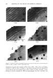
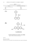
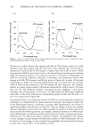
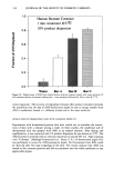
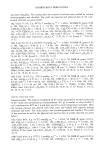
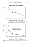
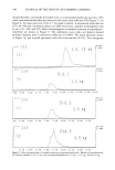
284 JOURNAL OF THE SOCIETY OF COSMETIC CHEMISTS _3. Own (a) (b) (c) (d) Figure 1. Scanning electron micrographs showing the surface of (a) untreated hair, and (b) DS-1, (c) DS-2, and (d) DS-3 hair. Refer to Table I for experimental conditions. layers are eliminated. The morphological change is discussed in detail in the succeeding section. CHANGE IN THE CROSS-SECTIONAL AREA OF CUTICLE CELLS TEM microphotographs of the cross section of the cuticle layers are represented in Figure 3 (a-f). Eight or nine layers of cuticle cells are clearly observed in the untreated hair depicted in Figure 3a. The black spots found in the cortical cells, and having an average diameter of 0.3 Ixm, are melanin pigment. One cuticle layer is constructed with two components that are differently stained. In Figure 3a, they are indicated as points A and B, which correspond to exocuticle and endocuticle, respectively (9). In DS-1 hair, the decrease in thickness of the outermost and subsequent three layers is detected. Besides, we can find a heavily stained layer (indicated by C in Figure 3b) that is not observed in the untreated hair. The boundary between exo- and endocuticles is getting obscure. These findings suggest that the reagent affects the chemical structure and that some amino acid components are dissolved even on the inner side of the cuticle, resulting in the loosely packed structure. However, the number of the layer is kept unchanged between untreated hair and DS-1, while in DS-2, as shown in Figure 3c, the number of layers diminishes to about five. The boundary between exo- and endocuticles in the
Purchased for the exclusive use of nofirst nolast (unknown)
From: SCC Media Library & Resource Center (library.scconline.org)