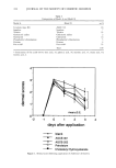
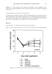
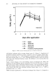
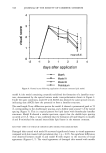
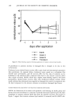
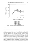
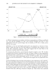
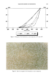
j. Soc. Cosmet. Chem., 47, 337-349 (November/December 1996) Measuring the encapsulation of cosmetic ingredients into liposomes: A method for large, hydrophilic compounds DELPHINE IMBERT,* GERALD B. KASTING, and R. RANDALL WICKETT, The University of Cincinnati College of Pharmacy, Division of Pharmaceutical Sciences, 3223 Eden Avenue, Cincinnati, OH 45267-0004 (D.I., R.R.W.), and The Procter & Gamble Company, Miami Valley Laboratories, Cincinnati, OH 45253-8707 (G.B.K.). Presented in part at the 1995 AAPS Annual Meeting, Miami Beach, and in part at the 1996 SCC Annual Scientific Seminar, Boston. Synopsis Encapsulation efficiency (EE) is an important parameter for the characterization of liposome systems en- trapping hydrophilic compounds. In this study, we were interested in quantifying EE of inulin, a 5,000-Da polysaccharide molecule, in various phospholipid liposome systems. Methods to separate free from encap- sulated inulin were evaluated. Dialysis and the centrifuged-column method were not appropriate for our liposome systems. Conversely, the Ficoll © discontinuous density gradient method was found to be an excellent tool for separating inulin encapsulated in large, multilamellar liposome preparations from free inulin. Using this method, we found that EE values approaching 40% were obtained by the reverse-phase evaporation method, regardless of cholesterol level. The microstructure was shown to consist of large heterogeneous multilamellar vesicles with diameters of 2-20 lam. At equal lipid concentration, much lower EE values (ca. 4%) were obtained for liposomes prepared by the film method. For inulin, comparison with other studies shows that EEs should be measured rather than inferred from systems having different compositions or preparation methods. INTRODUCTION Several investigations in animal models in vitro (1) and in vivo (2) have suggested that liposomal encapsulation can enhance the topical delivery of large hydrophilic com- pounds. In liposome preparations, these compounds may be located either in the aqueous core of the vesicles (encapsulated compound) or in the external medium (free compound). Hence, the extent of entrapment (encapsulation efficiency, EE) is a key parameter for the liposome formulator. Despite the widespread use of liposomes in cosmetic products, only a few research articles on this topic are available to the cosmetic scientist (3-5). Beyond formulation characterization, knowledge of EE in topical liposome testing also allows one to assess whether incorporation of the ingredient of interest into the liposome is important in altering distribution in the skin. * Current address: UCSF School of Pharmacy, Department of Biopharmaceutical Sciences, Box 0446, 513 Parnassus Avenue, San Francisco, CA 94143-0446 email: imbert@rebusucsf. edu. 337

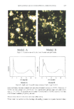
338 JOURNAL OF THE SOCIETY OF COSMETIC CHEMISTS EE is typically determined by separating free from encapsulated compound and assaying one or both fractions for the compound of interest. Various separation techniques have been described and compared in the literature. Current procedures utilize size exclusion gel filtration (6-12), centrifuged size exclusion gel mini-column (13-15), dialysis (16,17), ion exchange columns (18), ultracentrifugation (6,19-21), and discontinuous density gradients (22,23). For the encapsulation of hydrophilic molecules, typically, small-size vesicles (400 nm in diameter) encapsulating sucrose or carboxyfiuorescein have been evaluated (3,5,13,22,24). Inulin is a 5,000-Da hydrophilic molecule that has been reported to be localized in the skin following delivery from liposomes (1,25,26). We were interested in quantitating the EE of inulin in different liposome systems for i, vitro skin penetration experiments (27). Using a phosphatidylcholine (PC)/inulin liposome system, we tested the centri- fuged-column method, dialysis, and the discontinuous density gradient separation method. After selecting the latter as the optimum separation method for this particular system, we evaluated various encapsulation procedures and liposome compositions. MATERIALS AND METHODS CHEMICALS Soybean phosphatidylcholine (PC) and phosphatidylethanolamine (PE) were obtained from Avanti Polar Lipids (Alabaster, AL). Inulin, cholesterol, cholesteryl hemisuccinate (CHEMS), dicetylphosphate (DCP), oleic acid (OA), o•-tocopherol, N-2- hydroxyethylpiperazine-N'-2-ethanesulfonic acid (HEPES), and disodium EDTA were purchased from Sigma (St. Louis, MO). [Carboxyl-•4C]inulin (•4C-Carb-I, specific ac- tivity 2.05 mCi/g) was purchased from American Radiolabeled Chemicals (St Louis, MO), and L-3-phosphatidylcholine 1,2-di[1-•4C]palmitoyl (•4C-DPPC, specific activity 100-120 mCi/mmol) was obtained from Amersham (Arlington Heights, IL). Ficoll © 400-DL and Sephadex G-25 fine and G-50 fine were purchased from Pharmacia Biotech (Piscataway, NJ). The scintillation cocktail was Scintisafe Plus 50% (Fisher Scientific, Pittsburgh, PA). All other chemicals were of analytical grade. The buffer was HEPES buffer, pH 7.4 (20 mM HEPES, 150 mM NaC1, 0.1 mM EDTA). PREPARATION OF •4C-DPPC LIPOSOMES PC liposomes labeled with •4C-DPPC were prepared by the film method (28). Briefly, 30 mg PC, 1.65 pg •4C-DPPC (0.25 pCi), and 0.3 mg o•-tocopherol in chloroform solution were pipetted into a 100-ml round-bottom flask. The flask was mounted onto a rotary evaporator and the chloroform evaporated under a gentle N 2 stream. Once a dry lipid film was obtained, vacuum was applied for one hour to remove any traces of solvent. The film was then hydrated at room temperature for one hour with 5 ml buffer. The final PC concentration was 6 mg/ml, and the 14C-DPPC radiochemical concentra- tion was 0.05 pCi/ml. PREPARATION OF •4C-CARB-I LIPOSOMES Liposome preparations containing 2% w:v inulin spiked with •4C-Carb-I were prepared by four different methods. For the film method (28), 48 mg PC and 0.48 mg o•-to-
Purchased for the exclusive use of nofirst nolast (unknown)
From: SCC Media Library & Resource Center (library.scconline.org)