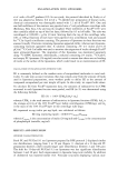
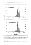
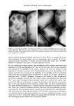
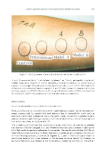
ENCAPSULATION INTO LIPOSOMES 339 copherol in chloroform solution were pipetted into a 25-mm x 175-mm test tube with a 24/40 ground-glass joint. The test tube was mounted onto a rotary evaporator, and a dry lipid film was obtained as described above. The film was then hydrated at room temperature for one hour with 2 ml buffer containing 2% w:v inulin and 0.14 pCi/ml •4C-Carb-I. The liposome preparation was submitted to five freeze/thaw cycles (isopro- panol-dry ice/25øC-water bath for 5 min). The final lipid concentration was 24 mg/ml. Three variations of the reverse phase evaporation method (RPE method) originally described by Szoka et M. (29) were studied. In protocols i and 2, 48 mg ofphospholipids, o•-tocopherol, and in some cases, other liposome wall components in chloroform solution were pipetted into a 25-mm x 175-mm test tube with a 24/40 ground-glass joint (22,24). The test tube was mounted onto a rotary evaporator and the chloroform evap- orated under a gentle N 2 stream. In protocol 1, once a dry lipid film was obtained, 6 ml of diethyl ether and 2 ml of buffer containing 2% w:v inulin and 0.14 pCi/ml •4C- Carb-I were added to the test tube. In protocol 2, 2 ml of diethyl ether and 0.67 ml of buffer containing 6% w:v inulin and 0.42 pCi/ml •4C-Carb-I were used. In both cases, the ether/buffer mixtures were sonicated for about 10-15s at 0-5øC to form a homo- geneous dispersion. Diethyl ether was then carefully removed under vacuum. In protocol 2, liposome preparations so obtained were then diluted 1:2 v:v with buffer, before use. The final liposome concentration was 24 mg/ml. For protocol 1, other concentrations were also studied. In the third RPE method (6), chloroform was used directly as the organic solvent instead of diethyl ether, and cholesterol and dicetylphosphate (DCP) were added as stabilizers. In a 100-ml round-bottom flask were dissolved 10.2 mg PC, 5.0 mg cholesterol, and 2.0 mg DCP, with 4 ml of chloroform. The flask was hand-shaken to dissolve all compo- nents, and 1 ml of buffer containing 2% w:v inulin and 0.14 pCi/ml 14C-Carb-I was added drop-wise. Chloroform was removed under vacuum. By this method, Alam et M. were able to encapsulate gelonin (a 30,000-Da protein) at a 77-79% efficiency (6). All liposome preparations were stored at 4øC under nitrogen before use. LIPOSOME SIZING Size distributions for PC and PC/Chol 9:1 mr liposomes prepared by RPE protocol 2 were determined using a HORIBA LA-900 laser scattering particle size analyzer (Horiba Instruments, Inc., Irvine, CA). The instrument combines QELS and Fraunhofer diffrac- tion to cover a range from -20 nm to -100 pm. Measurements were taken without sonication, and the refractive index was set at 1.19-0.00 i. CRYO-TRANSMISSION ELECTRON MICROSCOPY A PC liposome preparation prepared by RPE protocol 2 was examined by cryo- transmission electron microscopy (cryo-TEM) to characterize the microstructure. A 5-pl drop of liposomal dispersion was applied to a lacey carbon Formvar film-covered EM grid (Ted Pella, Inc., Redding, CA) in a controlled environmental vitrification system (30). Most of the solution was blotted from the grid with filter paper (Whatman •1) to form a thin film specimen, which was immediately vitrified by plunging into liquid ethane at its freezing point. The vitrified sample was examined at -172øC using a Gatan

340 JOURNAL OF THE SOCIETY OF COSMETIC CHEMISTS cold stage (Gatan, Inc., Warrendale, PA) in a Philips CM-12 TEM (Philips Electronic Instruments Co., Mahwah, NJ). Acceleration voltage was 100 kV. Micrographs were recorded on Kodak SO-163 film developed to maximum speed in full-strength Kodak D- 19 developer. CENTRIFUGED MINI-COLUMN SEPARATION METHOD The centrifuged mini-column method is a combination of Sephadex filtration and low- speed centrifugation, which prevents sample dilution typically observed in conventional size-exclusion gel filtration. It is non-destructive, fast (about 5 min/sample), and uses sample sizes in the 0.1-0.5 ml range. Free inulin is retained by the gel whereas lipo- somes are recovered in the eluate. The methods of Fry et al. (13) and Penefsky (15) were adapted as follows: Sephadex beads were allowed to swell in buffer. The barrel of a 3-cc syringe (Becton-Dickinson, Rutherford, NJ), fitted with drain disks (Corning Costar Corp., Boston, MA) was filled with Sephadex gel and inserted into a 15-ml conical centrifuge tube, supported at the top by the finger grips of the syringe. The whole assembly (syringe and tube) was centrifuged for two minutes at 100 g to remove excess buffer from the Sephadex beads. The syringe was transferred to a clean tube, and 0.5 ml of the test preparation was carefully applied to the Sephadex bead. The column was centrifuged as before and the eluate recovered for liquid scintillation analysis. Two grades of Sephadex, Sephadex G-50 fine and G-25 fine, were evaluated. Retention of free inulin by the Sephadex gel was tested using a solution of 2% w:v inulin and 0.14 laCi/ml •4C-Carb-I in buffer. Liposome recovery from the column was assessed with a •4C-DPPC liposome preparation (0.05 laCi/ml). Radioactivity levels in the eluates were determined by scintillation counting on a Beckman LS 7000 scintillation counter (Beck- man Instruments Inc., Irvine, CA). DIALYSIS The dialysis protocol described by Bangham et al. (16) was modified to account for the difference in diffusion rate between small ions and inulin. Dialysis tubing (Fisher Scientific, Pittsburgh, PA cut-off, 12,000 Da) was cleaned according to a standard procedure and rinsed with buffer. The lower end of the tube was tightly clipped, and 0.5-1.0 ml of the test solution was pipetted into the tube. The upper end was clipped tight and the sample dialyzed against 12-hour changes of 500 ml buffer. A solution of 2% w:v inulin and 0.14 laCi/ml •4C-Carb-I in buffer and •4C-DPPC liposomes was tested. Radioactivity levels in the dialysates and dialysis bag were determined by scin- tillation counting. DISCONTINUOUS FICOLL © DENSITY GRADIENT SEPARATION METHOD In the discontinuous density gradient method, layers (typically three or four in a 5-ml centrifuge tube) of decreasing density are carefully pipetted on top of each other, with the sample mixed in the layer of highest density. Upon centrifugation, separation occurs based on molecular weight. This method was used successfully by Betageri with a metrizamide gradient (31), by Heath et al. with a dextran gradient (23), and by Fraley
Purchased for the exclusive use of nofirst nolast (unknown)
From: SCC Media Library & Resource Center (library.scconline.org)