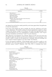
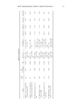
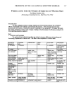
82 JOURNAL OF COSMETIC SCIENCE Table II Composition of O/W Emulsion Ingredient % W/W Benzophenone-3 Octyl p-methoxycinnamate Diisopropyl adipate DEA-cetyl phosphate Polymer (if present) Propylene glycol (and) diazolidinyl urea (and) methyl paraben (and) propyl paraben Distilled water qs with distilled water to 6.0 7.5 8.5 2.0 5.0 1.0 65.3 100 was drained out and the skin surface patted dry with tissue paper before the application of sunscreen preparations. Finite dosing. Finite dosing was used to simulate the actual use conditions in all of the in vitro experiments. The smallest volume of sunscreen preparation required to obtain complete and uniform coverage of the diffusion cell surface area (approximately 0.636 cm 2) was determined to be 4 }al, corresponding to a weight of between 3 and 4 mg for prototype formulations and between 5 and 6.5 mg for emulsions. After application, the preparation was uniformly spread on the SC side of the skin with the help of a glass rod, and the tip of the rod was washed into a vial containing 2 ml of alcohol in order to account for the material lost in spreading. With this technique the exact amount of material applied to the skin surface was obtained. In vitro skin permeation methodology. A flow-through system was used to conduct in vitro experiments. The total system consisted of a receptor fluid reservoir a variable flow rate peristaltic pump, Cassette © (Manostat, New York, NY) a circulating water bath, Table III Composition of W/O Emulsion Ingredient % W/W Benzophenone-3 6.0 Octyl p-methoxycinnamate 7.5 Polyglycerol-3-DI-IS 11.0 Beeswax 0.5 Mineral oil 5.0 Octyl dodecyl myristate 4.0 Mineral oil (and) apricot kernel oil (and) calendula extract (oily vegetol marigold WL 1072) 4.5 Methyl paraben 0.1 Polymer (if present) 5.0 Glycerin 5.0 Sodium chloride 0.5 Magnesium sulfate 0.5 Deionized water 50.0 Carbomer 940 0.3 Imidazolidinyl urea 0.3 Triethanolamine 99% 0.3

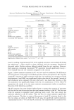
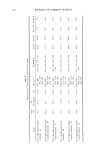
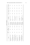
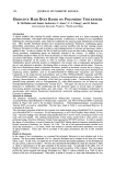
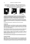
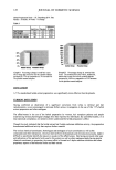
WATER RESISTANCE OF SUNSCREENS 83 Lauda © (Brickman Instrument, Westbury, NY) two cell-holding heating blocks 14 Teflon © flow-through diffusion cells and a Retriever IV fraction collector (ISCO Inc., Lincoln, NE) to collect effluent fractions over the selected time period. The skin surface temperature was maintained at 32øC by adjusting the circulating water bath tempera- ture to 39øC (8). The effluent from diffusion cells were collected directly into polyeth- ylene scintillation vials and held for analysis. The protocol included 20 minutes of sunscreen drying time after application to the skin, followed by 20 minutes of wetting with 1 ml of water, and then pouring the water out from the cells for analysis. These steps were repeated a second time. In total there were 40 minutes of drying and 40 minutes of water exposure. In each experiment a minimum of four replicates was used. TISSUE SAMPLE PREPARATION AND ANALYSIS The liquid scintillation counting technique was used to analyze all the i, vitro study samples. At the conclusion of the experiment, scintillation fluid was added to the effluent samples collected directly into the vials and the amount of sunscreens penetrated was estimated from the counts of radioactivity present in the samples. The skin was removed from the cell, and the stratum corneum was separated from the rest of the epidermis to obtain the amount of material remaining inside the barrier layer of the skin by tape-stripping. In this technique, seventeen strippings of the sunscreen-treated site of the skin using transparent 3M Scotch TM tape were taken on a single piece. For each strip, a fresh tape surface was used. Sunscreens recovered from the 17 strips were considered as the amounts inside the SC. Scintillation fluid was added to the vials containing tape strips and allowed to stand at room temperature for at least 24 hours to enable extraction of sunscreens in order to perform scintillation counting on the samples. The remainder of the skin was digested in 2 ml of tissue solubilizer at 50øC for 24 hours in an oven. This was done to get the estimate of material in the viable tissues of the skin. Digested skin samples were neutralized with glacial acetic acid, and scintillation fluid was added for scintillation counting. Thus the amount of sunscreen was estimated in four locations in each in vitro experiment: (i) the receptor fluid, (ii) the washes, (iii) SC (from the strippings), and (iv) viable tissues of the skin (from the digested skin). Sigma Stat 2.0 (Jandel Scientific, San Rafael, CA) was used to perform statistical analyses of the data. Whenever analysis of variance of data showed significant differences among observations, Dunnett's test was used, as it allowed comparisons with the control ob- servation. RESULTS AND DISCUSSION PROTOTYPE FORMULATIONS Tables IV and V show sunscreen washoff and skin distribution for the simple prototypes. Recovery of the labels was excellent, usually between 85% and 105% of the theoretical values. The washoff data for the controls (no polymer present) are remarkably consistent for both sunscreens and both formulations. Between 50% and 60% of each sunscreen was removed by water washing under the conditions employed. Significant quantities were
Purchased for the exclusive use of nofirst nolast (unknown)
From: SCC Media Library & Resource Center (library.scconline.org)