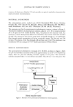
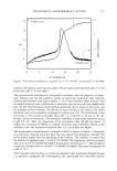
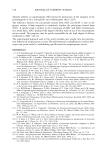
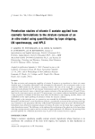
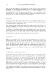
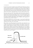
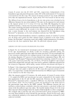
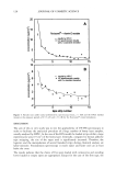
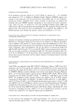
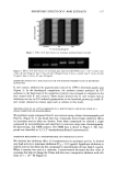
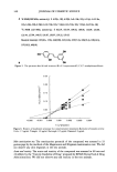
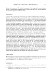
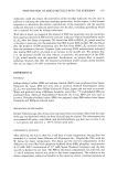
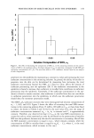
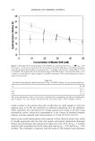
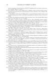
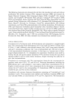
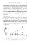
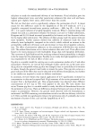
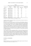
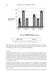
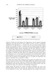
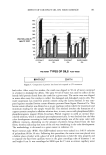
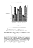
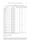
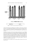
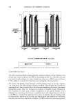
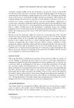
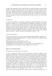
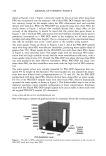
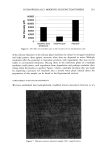
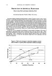
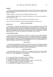
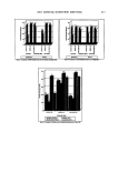
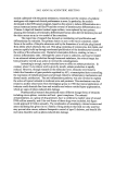
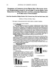
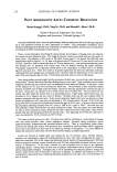
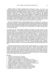
PENETRATION OF MIXED MICELLES INTO THE EPIDERMIS 149 •5 1.00 0.83 0.50 Solution Composition of SDS, as Figure 1. The effect of decreasing the composition of SDS, %, in the contacting solution on the concen- tration of SDS in the epidermis after a five-hour exposure (Cj/•,,,) to solutions containing 50 mM SDS and increasing concentrations of C12E6 . The error bars reflect a 95% confidence interval based on six samples of each composition. penetrate into the epidermis by maintaining a constant o• s value and increasing the total surfactant concentration in the contacting solution. In general, the ability of micelies to penetrate into the skin can be determined by measuring how increasing the total surfactant concentration beyond the CMC, at a fixed (x s value, affects the amount of surfactant penetrating into the epidermis (28). If the surfactant concentration in the epidermis is found to increase, then surfactant in miceliar form contributes to surfactant penetration into the epidermis. Conversely, if the surfactant concentration in the epi- dermis is found to remain constant, then surfactant in miceliar form does not contribute to surfactant penetration into the epidermis, in which case the surfactant penetration should obey the monomer penetration model. The SDS/C•2E 6 surfactant mixtures that were investigated had solution compositions of % = 1, 0.83, and 0.50. Figure 2 shows the effect of increasing the total SDS concen- tration in the contacting solution (from 25 mM to 100 mM) on C,,•i , at these three fixed % values. As shown in Tables I and II (30,31), for each value of % over the range of surfactant concentrations examined, %n = O•s, O• is constant and C• is approximately constant. Therefore, any observed increase in C.,3.i, as the total SDS concentration in- creases for each % value examined can only be attributed to the penetration of miceIlar SDS into the epidermis, because only the micelle concentration is increasing. (Recall that the SDS monomer concentration is equal to oqC•, which remains constant, while the concentration of SDS in miceIlar form is equal to %n(Ct - C•), where C• is the total surfactant concentration, which increases in this case.)

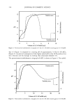
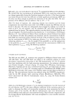
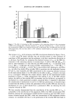
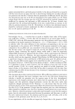
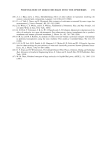
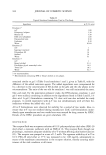
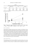
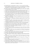
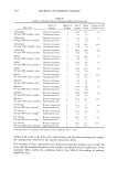
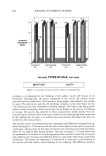
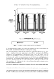
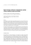
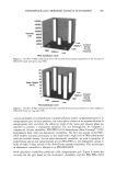
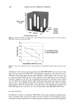
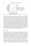
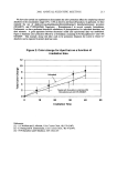
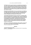
150 JOURNAL OF COSMETIC SCIENCE 9 8 7 .._,6 2 1 0 I 'I- 1.00 0.83 0.50 Solution Composition of SDS, =s Figure 2. The effect of increasing the SDS concentration in the contacting solution on the concentration of SDS in the epidermis after a five-hour exposure (C.,,•i,). For each composition (% = 1, 0.83, and 0.50), the concentrations of SDS in the contacting solution are 25 mM (empty bats), 50 mM (solid bars), and 100 mM (striped bars). The error bars reflect a 95% confidence interval based on six samples at each SDS concentration. The increase in Cski, , with increasing total SDS concentration observed in Figure 2 for tx• -- 1, 0.83, and 0.50 clearly indicates that the micelies present in these solutions do contribute to SDS penetration into the epidermis, with their contribution decreasing as tx s decreases. Specifically, by comparing the observed increase in Cski, , as the SDS con- centration in the contacting solution is increased from 25 mM to 100 mM (AC,•i, ,) for each tx s value examined, it is clear that the pure SDS micelies (ix s = 1) contribute more to C•i, , (AC•i, , • 0.08) than the mixed micelies corresponding to tx s = 0.83 (AC•in = 0.03) and to tx s = 0.50 (Ac,kin = 0.02). This is clear evidence that changing tXs, and hence tXm, can affect the ability of the miceliar SDS to penetrate into the epidermis, because for each tx s value examined, the SDS concentration in the contacting solution increases by the same amount (from 25 to 100 raM), but the effect on AC•i, , is found to decrease as tx s is decreased. Although this simple analysis, based on the experimental results presented in Figure 2, clearly demonstrates that adding C]2E 6 to the SDS solution reduces the ability of the miceliar SDS to penetrate into the epidermis, as proposed in mechanism (ii), a more quantitative analysis, presented below, is required to determine the contributions of mechanisms (i) and (ii) to SDS skin penetration. It should be kept in mind that the ability to reduce the penetration of the miceliar SDS into the skin by mechanisms (i), (ii), or both should have a pronounced effect on reducing the skin irritation induced by SDS. We have recently demonstrated that the contribution of the miceliar SDS to C•i, , is comparable to the contribution of the monomeric SDS at low SDS concentrations (28). However, because the concentration of SDS micelies increases as the total SDS concen- tration increases beyond the CMC, while the concentration of SDS monomers remains constant, we concluded (28) that it is the penetration of the miceliar SDS that leads to
Purchased for the exclusive use of nofirst nolast (unknown)
From: SCC Media Library & Resource Center (library.scconline.org)