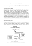
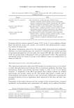
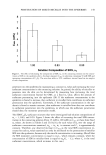
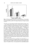
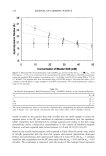
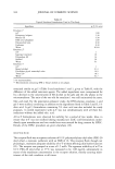
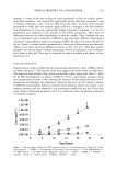
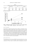
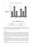
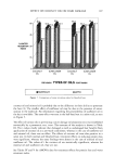
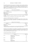
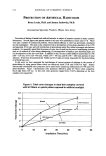
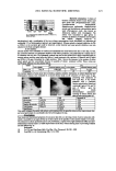
PENETRATION OF MIXED MICELLES INTO THE EPIDERMIS 151 the dose-dependent skin irritation response observed by many researchers (2,3,8,13,16,18). Indeed, we found that the SDS miceliar contribution overwhelms the SDS monomeric contribution at the higher SDS concentrations (28). Accordingly, re- ducing, or preventing, the contribution of the miceliar surfactant to C,/•i , by mixing surfactants should lead to a reduction in the skin irritation potential of the surfactant mixture, in addition to any beneficial effect due to a reduction in the surfactant mono- met concentration. REGRESSION ANALYSIS OF THE CONTRIBUTIONS OF MICELLAR AND MONOMERIC SDS TO Cs•,n FROM SOLUTIONS OF SDS/C•2E 6 Figure 2 shows that as o• s decreases, the contribution of the SDS/C•2E 6 mixed micelies to Cs•i, , decreases. To quantify the relative contributions of SDS in mixed micelle form (o• m = 1, 0.83, and 0.50) and in monomeric form to Cs•i,,, we carried out a multiple linear regression analysis using all the experimental data, prior to averaging, that was used to generate Figure 2. The simplest relationship between the SDS concentrations in miceliar and monomeric form to C•,•i,, is a linear one. The basis for this linear relationship is that in Fickian diffusion from an infinite reservoir with a large concentration differ- ence, the net permeant flux at a given time is directly proportional to the initial permeant concentration (42). With this assumption in mind, we fitted C•,•i,, to the following expression: C,•i, = o• ß C•,so s + b' C(o• m = 1) + c' C(o• m = 0.83) + d' C(o• m = 0.50) (4) where a, b, c, and d are the regression coefficients that were determined from the regression analysis, Cj,so s is the SDS monomer concentration, C(O•m) is the SDS con- centration in micelies of composition O•m, and C,/•, is the SDS concentration in the epidermis (in units of mmols of SDS per gram of dry epidermis). For the regression analysis, Cj,so s = oqC• and C(o•,•) = oq•(C t - Cj)using the appropriate values of o•, o•m, and Cj reported in Tables I and II (30,31). In this manner, we were able to isolate the contributions to C.,•i , due to the miceliar SDS for the three micelie compositions ex- amined (reflected in b, c, and d), as well as due to the monomeric SDS (reflected in a). The following values of a, b, c, and d were obtained from the regression analysis: 4.1 +_ 1.0 C,/•i,/Cl,so s 0.032 +0.014 Cskin/C(O• m '- 1) 0.003 +0.012 C•/•i,/C(o• m = 0.83) 0.0009 +_ 0.0092 C,/•i,/C(o• m = 0.50) According to these regression results, the O• m = 0.50 micelies do not contribute to C,/•, at all, because d is essentially equal to zero. The o• m = 0.83 micelies contribute very little or not at all to C,/•i ,, because although the average value of c is not zero, the 95% confidence interval includes zero. On a per SDS molecule basis, the contribution of the SDS monomers is quite large, with one SDS molecule in monomeric form being 130 times more skin penetrating than one SDS molecule in a pure SDS micelie (o• m = 1). However, at the higher SDS concentrations, there is significantly more miceliar SDS than monomeric SDS, and as a result, the net contribution to C•/•i , due to the miceliar SDS may overwhelm that due to the SDS monomers.

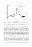
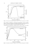
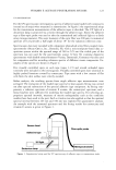
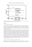
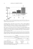
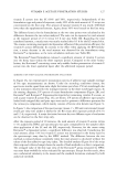
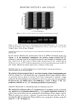
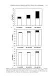
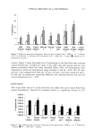
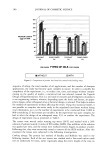
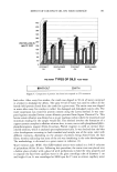
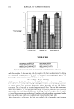
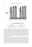
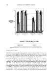
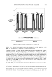
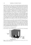
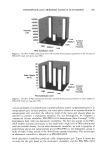
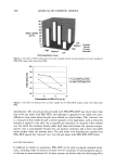
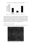
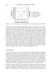
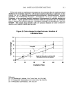
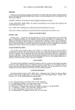
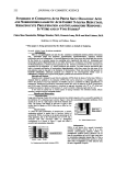
152 JOURNAL OF COSMETIC SCIENCE To more clearly describe the relative contributions of the monomeric SDS and the miceliar SDS to skin penetration, Figure 3(a-c) shows the total contributions of the monomeric and the miceliar fractions of SDS to Cs•i, for o• s = 1, 0.83, and 0.50 respectively, based on the regression data given above. Specifically, the SDS monomeric contribution to Cs•i, is a'C•,sos, and the three miceliar contributions to Cs•i, are b ß C(o• m = 1), c' C(o• m = 0.83), and d' C(o• m = 0.50). Figure 3(a-c) clearly shows that the SDS monomers make a contribution to Cs•i, that is larger than that of the miceliar SDS for the three o•s values examined, as seen by the empty bars (representing the monomeric contribution) always being larger than the solid bars (representing the miceliar contribution). It is only for o• = 1, the pure SDS case, that the micelies make a large contribution to Cs• , , particularly at the highest surfactant concentrations ex- amined (see Figure 3a). In Figures 3b and 3c, which correspond to the o• -- 0.83 and 0.50 surfactant mixtures, respectively, the miceliar contribution is almost non-existent. Indeed, considering the confidence interval for the coefficients c and d, it is apparent that the miceliar contributions include the possibility of a zero contribution to Cs• ,. There- fore, the monomer penetration model represents a reasonable approximation for the two SDS/C•2E 6 surfactant mixtures examined, where the miceliar SDS does not penetrate appreciably into the epidermis for the SDS concentrations examined (25, 50, and 100 raM). However, the reduction in Cs•i, observed with decreasing o• s, shown in Figures 1 and 2, results from both the reduction in the SDS monomer concentration and the reduction in the ability of the miceliar SDS to penetrate into the epidermis. Generalizing the observations made in the case of the SDS/C•2E 6 surfactant mixtures to other surfactant mixtures, it is plausible that the observed reduction in skin irritation upon mixing surfactants reported by several researchers occurs because both the toohomeric and the miceliar s•rfactant penetrations into the skin are diminished (6,24,26). At the high total surfactant concentrations commonly utilized in commercial surfactant products, the micellar contribution can be quite large, as demonstrated by the dose-dependent surfactant-induced skin irritation results reported in the literature (2,3,8,13,16,18). Consequently, any reduction in the ability of the micellar surfactant to penetrate into the skin, as reflected by lower values of the regression coefficients (such as b, c, and d), should have a significant impact on C_,• n at high total surfactant concentrations. In other words, reducing the miceliar contribution to Cs•i, should lead to a reduction in the skin irritation potential of the surfactant system contacting the skin. DYNAMIC LIGHT SCATTERING DETERMINATION OF SDS/C•2E 6 MIXED MICELLE SIZES In Figure 4, the hydrodynamic radii of the SDS/C•2E 6 mixed micelles are determined using DLS by extrapolating the effective hydrodynamic radii of these micelies to a zero micelie concentration. At the surfactant concentrations corresponding to Figure 4, O• m is predicted to be approximately equal to o•, and therefore, one can treat the micelies as having a constant O• m value over the entire surfactant concentration range examined (see Table I). This is important, because a change in o• m could lead to a change in the hydrodynamic radii of the micelies. (The hydrodynamic radii of the micelies determined using this method are reported in Table III.) According to the surfactant penetration model advanced in our recent paper (28), the size of the micelie determines its ability to penetrate into the SC. (Note that the discussion in the following section introduces the caveat that electrostatic interactions may also play a role.) The micelie penetration
Purchased for the exclusive use of nofirst nolast (unknown)
From: SCC Media Library & Resource Center (library.scconline.org)