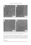
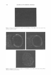
298 JOURNAL OF COSMETIC SCIENCE is composed of a tram-line structure including a densely stained 8-layer (proteins) sandwiched between lightly stained J3-layers (lipids) on both sides (1). It has been proposed that lipids of the CMC comprise a bilayer structure, based upon the fact that lipids extracted from wool or human hair are able to form liposomes (4,5). A strong orientation of hair lipids, probably at the CMC, in planes parallel to the axis of hair fibers, has been recently shown using a microbeam synchrotron radiation diffraction technique (6). Therefore, it has been suggested that lipids of the CMC play roles in the physicochemical phenomena of hair fibers, such as chemical diffusion, cell cohesion, and mechanical strength (2,3,7-9). Since the CMC has a thickness of approximately 25 nm (1-3), optical microscopy is not able to resolve the CMC structure. On the contrary, as transmission electron microscopy (TEM) has conventionally been used to observe the CMC (1-3), electron microscopy (which is superior in spatial resolution to optical microscopy) provides sufficient reso lution to analyze the subtle structures of hair fibers. However, the J3-layers in TEM images always appear as negative staining patterns, not attributable to the existence of the lipids, which implies that the J3-layers do not directly reflect lipids distributed in the CMC. Although scanning electron microscopy (SEM) can clarify the fine structure of hair fibers, it is impossible to observe lipids in the CMC using SEM, even of a polished hair plane, since detection in SEM is based upon secondary electrons derived from an uneven surface. Thus, there are currently no available methods to directly visualize lipids in the CMC of hair fibers. We have recently found that argon sputter etching-SEM (ASE-SEM) of a transversely polished hair plane provides a specific characteristic image, especially at the cortex. This finding prompted us to determine how the characteristic image on the surface of the hair plane is generated by ASE-SEM and to validate the use of ASE-SEM images in research on the microstructures of hair fibers. In this study, we have characterized ASE-SEM images of the hair plane by examining the effects of several treatments on the ASE-SEM images. MATERIALS AND METHODS CHEMICALS Epon 812 resin, 25% glutaraldehyde, and osmium tetroxide were from TAAB (Reading, UK). Oleyl oleate, myristyl myristate, and isopropyl palmitate were from Kao (Tokyo, Japan), while other fats and oils were from Tokyo-Kasei (Tokyo, Japan) or Wako (Tokyo, Japan). MATERIALS Scalp hair fibers were obtained from a healthy Japanese volunteer, aged 28 years. The volunteer had never subjected her hair to any chemical treatments such as perming or coloring, except for shampooing and conditioning. The hair fibers were washed with n-hexane for 5 min and then were cut to ca. 1 cm prior to preparation for ASE-SEM.

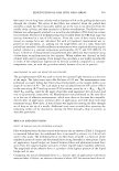
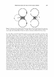
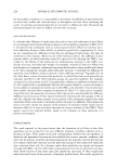
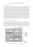
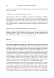
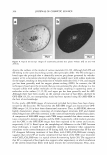
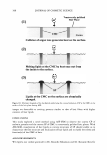
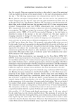
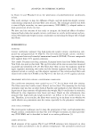
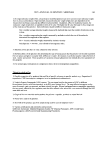
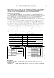
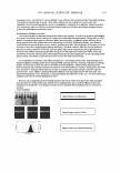
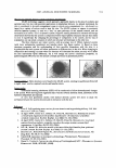
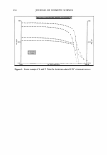
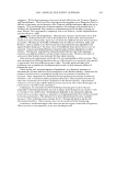
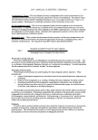
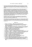
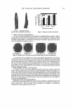
VISUALIZING HAIR LIPIDS BY ASE-SEM 299 ASE-SEM OBSERVATION The 1-cm hair fiber was embedded in resin, and then a transversely polished resin embedded hair block was exposed to ASE for SEM observation. The Epon 812 resin containing the hair fiber was polymerized in an oven. Rough trimming of each hair block with a glass knife was followed by the careful polishing of a transverse hair plane with a diamond knife (MT6740, DiATOME Ltd., Switzerland). The carefully polished hair block was placed in the target position for ASE in an Hitachi Ion Sputter E-1030 (Hitachinaka, Japan) connected to an argon cylinder. The specimen was then etched under a pressure of 6 to 7 Pa, a current of 6 mA, a voltage of 0.3 kV, a working distance of 30 mm, and a time of 360 sec, as shown in Figure 1. The etched specimen was coated with a ca. 5-nm layer of Pt-Pd using the same instrument (Hitachi Ion Sputter E-1030). Observation of the polished hair plane was performed using an FE-SEM Hitachi S-4300 SEM under an accelerating voltage of 5 kV. For observation of thin sections, 1-µm-thick sections were cut with a sapphire knife while 90-nm ultra-thin sections were cut with a diamond knife. ASE was then performed for the hair sections on a cover glass using the same procedures and the same conditions as those used for the polished hair planes. COMPARISON OF ASE-SEM IMAGES WITH TEM IMAGES The transversely polished resin-embedded hair block was prepared according to the procedures described above. An ultra-thin section of the block was cut with a diamond knife, and then was stained with uranyl acetate and lead citrate (3). The ultra-thin sections were observed with a TEM (Hitach H-7000) under an accelerating voltage of 75 kV. If a clear TEM image was obtained, another polished hair plane of the remaining block was examined by ASE-SEM. In case of an unclear TEM image, further ultra-thin Electrode rgon tm phere 6-7P 360 30mm Target 6mA 0.3 kV Figure 1. Schematic diagram of the instrument used for ASE in this study.
Purchased for the exclusive use of nofirst nolast (unknown)
From: SCC Media Library & Resource Center (library.scconline.org)