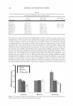
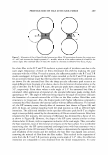
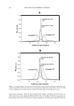
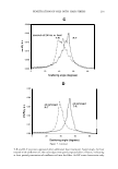
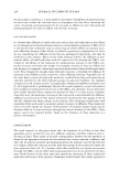
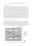
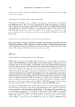
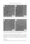
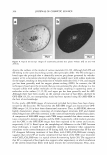
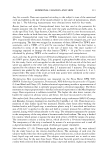
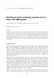
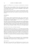
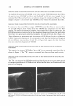
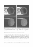
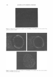
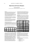
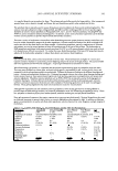
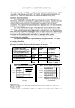
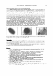
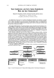
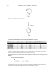
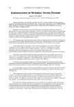
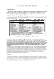
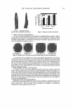
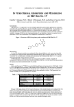
300 JOURNAL OF COSMETIC SCIENCE sections were cut and subsequent TEM observations were continued until a clear TEM image were recorded. TREATMENTS OF HAIR FIBERS AND POLISHED HAIR PLANES Treatments of hair fibers were as follows: (a) immersion of hair fibers into CH 3 Cl/ CH�OH/water (v/v/v, 18:9: 1) at room temperature for 24 hr, (b) immersion of solvent treated hair fibers into melting lipids at 80°C for 24 hr, and (c) chemical fixation with glutaraldehyde and osmium tetroxide. The polished hair planes were observed with ASE-SEM followed by pre- and post-incubation with ethanol or n-hexane. Pre- and post-incubations refer to incubations with the solvent just before ASE and just after ASE, respectively. OBSERVATION OF THE POLISHED HAIR PLANE BY OPTICAL MICROSCOPY Optical microscopic images of polished hair planes were observed with a Microwatcher (Mitsubishi Chemical, Tokyo, Japan) via a reflection mode. The IR (infrared) spectrum of the polished hair plane was measured with an IR microscope (Spectrum One, Perkin Elmer, UK) using an ATR (attenuated total reflection) mode. RESULTS CHARACTERISTICS OF ASE-SEM IMAGES OF THE HAIR PLANE SEM images of transversely polished hair planes with or without ASE are shown in Figure 2. In the SEM images without ASE, as used conventionally, the hair plane can be visualized only as the even surface, and therefore, micro-three-dimensional structures of the cuticle and the cortex in the hair plane could not be observed (Figure 2a). In contrast, when ASE treatment was performed on the hair plane prior to SEM observation, mi crostructures of the cuticle and the cortex became distinct with a characteristic pattern at the cortex (Figure 26). The magnified image of the cortex revealed that it was mainly composed of two types of structure, circular-shaped ones and slender thread-shaped ones, designated here as circular patterns (CP) and stitch patterns (SP), respectively. When several parameters associated with the efficiency of ASE, such as pressure, voltage, current, working distance, and treatment time, were characterized, we found that of all parameters tested, pressure most strongly affected ASE-SEM images of the hair plane, and that a pressure of 6 to 7 Pa is required for an optimal ASE-SEM image. A comparison of ASE-SEM images at the tilting angle of 45 degrees toward the primary electron beam with images at no tilting angle demonstrated that both CP and SP are convex structures in the surrounding plane (Figure 3). In order to determine the intra or intercellular localization of CP and SP in the cortex, TEM images of ultra-thin sections and ASE-SEM images in the adjacent plane were observed. In a representative TEM image (Figure 4a), there are densely stained melanin granules and the lightly stained CMC. A comparison of each TEM image with the adjacent ASE-SEM image

VISUALIZING HAIR LIPIDS BY ASE-SEM 301 Figure 2. SEM images of transversely polished hair planes without ASE (a) and with ASE (b). ASE-SEM image (b) includes a circular pattern (CP) and a stitch pattern (SP) in the magnified image of the cortex. Cu: cuticle. Co: cortex.
Purchased for the exclusive use of nofirst nolast (unknown)
From: SCC Media Library & Resource Center (library.scconline.org)