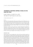
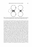
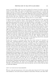
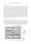
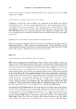
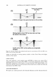
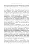
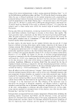
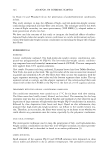
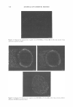
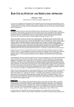
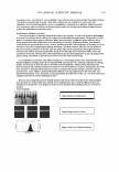
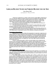
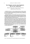
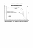
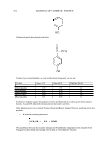
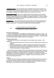
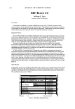
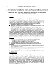
308 (1) JOURNAL OF COSMETIC SCIENCE Transversely polished Hair Plane Collision of argon ions generates heat on the surface. (2) Melting lipids at the CMC by heat ooze out from the inside to the surface. (3) Lipids at the CMC on the surface are chemically changed. Figure 10. Schematic diagram of the mechanism underlying the convex formation of SP at the CMC on the surface of the hair plane during ASE. images are detectable as staining patterns similar to that of hair fibers with higher contents of hair lipids. CONCLUSIONS This study explored a novel method using ASE-SEM to observe the convex CP of melanin granules and the convex SP of CMC in transversely polished hair planes. With ASE-SEM, visualization of hair lipids as convex structures of SP should enable us to characterize the fine structure and localization of hair lipids and to clarify the role(s) and function(s) of the CMC of hair. ACKNOWLEDGMENTS We express our cordial gratitude to Dr. Kouichi Nakamura and Dr. Katsumi Kita for

VISUALIZING HAIR LIPIDS BY ASE-SEM 309 their invaluable discussions and guidance in this study. Our sincere thanks are also due to Ms. Miho Suzuki for technical support in the SEM studies. REFERENCES (1) J. A. Swift and A. W. Holmes, Degradation of human hair by papain. Part II. Some electron micro scope observations, Text. Res.]., 35, 1014-1019 (1965). (2) J. D. Leeder, The cell membrane complex and its influence on the properties of the wool fibre, Wool Sci. Rev., 63, 3-35 (1986). (3) S. Naito, T. Takahashi, M. Hattori, and K. Arai, Histochemical observation of the cell membrane complex of hair, Sen-I Gakkaishi, 48, 420---426 (1992). (4) A. Korner, S. Petrovic, and H. Hocker, Cell membrane lipids of wool and human form liposomes, Text. Res.]., 65, 56-58 (1995). (5) L. Coderch, A. de la Maza, A. Pinazo, and J. L. Parra, Physicochemical characteristics of liposomes formed with internal wool lipids,]. Am. Oil Chem. Soc., 73, 1713-1718 (1996). (6) L. Bertrand, J. Doucet, A. Simionovici, G. Tsoucaris, and P. Walter, Lead-revealed lipid organization in human hair, Biochim. Biophys. Acta, 1620, 218-224 (2003). (7) K. Nishimura, M. Nishina, Y. Inaoka, Y. Kitada, and M. Fukushima, Interrelationship between the hair lipids and the hair moisture,]. Cosmet. Sci. Soc. Jap., 13, 134-139 (1989). (8) V. Sideris, L.A. Holt, and I. H. Leaver, A microscopical study of the pathway for diffusion of rhodamine B and octadecylrhodamine B into wool fibers,]. Soc. Dyers Colorists, 106, 131-135 (1990). (9) M. Philippe, J.C. Garson, P. Gilard, M. Hocquaux, G. Bussler, F. Leroy, C. Mahieu, D. Semeria, and G. Vanlerberghe, Synthesis of 2-N-oleoylamino-octadecane-1,3-diol: A new ceramide highly effective for the treatment of skin and hair, Int. J. Cosmet. Sci., 17, 133-146 (1995). (10) R. S. Thomas and J. R. Hallahan, Use of chemically-reactive gas plasmas in preparing specimens for scanning electron microscopy and electron probe microanalysis, Scanning Electron Microsc., l, 83-92 (1974). (11) T. Fujita, T. Nagatani, and A. Hattori, A simple method of ion-etching for biological materials. An application to blood cells and spermatozoa, Arch. Histol. Jap., 36, 195-204 (1974). (12) T. Nagatani and M. Yamada, Application of ion-etching for biological materials, The Cell (Tokyo), 7, 136-142 (1975). (13) W. J. Humphreys and W. G. Henk, Ultrastructure of cell organelles by scanning electron microscopy of thick sections surface-etched by an oxygen plasma,]. Microsc., 116, 255-264 (1979). (14) J. A. Swift, A technique for the rapid examination of the gross internal structure of mammalian keratin fibres,]. Text. Inst., 3, 170-174 (1980). (15) S. Seta, H. Sato, M. Yoshino, and S. Miyasaka, SEM/EDX analysis of inorganic elements in human scalp hairs with special reference to the variation with different locations on the head, Scanning Electron Microsc., l, 127-140 (1982). (16) R. Sellamuthu, J. Barkanic, E. Karwacki, and R. Jaccodine, Application of SEM and XPS in plasma etching of single crystal silicon, Microbeam Anal., 21, 65 3-65 5 (1986). (17) A. C.-M. Yang, R. D. Allen, and T. C. Reiley, Evaluation of particle dispersion in polymer solids by oxygen plasma etching,]. Appl. Polym. Sci., 46, 757-762 (1992). (18) G. Danev, E. Spassova, and K. Popova, Morphology of thin polyimide films, Thin Solid Films, 228, 301-303 (1993). (19) T. Nagata, Y. Hisanaga, D. Shimizu, and T. Fujiwara, Scanning electron microscopy of ion-etched epoxy resin sections and its application to three-dimensional reconstruction, Electron Microsc., 36, 131-134 (2001). (20) E. G. Bligh and W. J. Dyer, A rapid method of total lipid extraction and purification, Can. J. Biochem. Physiol., 37, 911-917 (1959).
Purchased for the exclusive use of nofirst nolast (unknown)
From: SCC Media Library & Resource Center (library.scconline.org)