324 JOURNAL OF COSMETIC SCIENCE by Chow (3) and Woodard (4) on the penetration of polyethylenimine conditioners in hair. This study attempts to map the diffusion of high- and low-molecular-weight cationic conditioning compounds into hair-fiber cross sections. The technique used for this work is time-of-flight secondary ion mass spectrometry (TOF SIMS) (5 ), adapted earlier to show penetration of oils into hair. We have used the outcome of this study to interpret the beneficial effects of surface deposited high-molecular-weight cationic conditioner on cuticle reinforcement and pen etrated low-molecular-weight cationic conditioner on extending the fatigue life of single hair-fibers. EXPERIMENT AL MATERIALS Cationic conditioning compounds. The high-molecular-weight cationic conditioning com pound was polyquaternium-10 (PQ-10). The low-molecular-weight cationic condition ing compound was cetyl trimethyl ammonium bromide (CETAB). The neat compounds were applied from 0.5% aqueous solutions. Hair samples. Fourteen-inch-long unaltered, European brown hair from DeMeo Brothers, New York, was used in this work. The top five inches of the root sections were mounted in parallel and identified as #1-20. The fibers were then cut into two segments, with the upper segments measuring two inches and the bottom segments three inches. The top segments served as controls, and the adjacent segments of the corresponding fibers were treated with either the CETAB or the PQ-10 in the form of a 0.5% aqueous solution. TREATMENTS WITH THE CATIONIC CONDITIONING COMPOUNDS The conditioner treatments were carried out at 3 7 ° C for six hours with slow stirring. The fibers were then briefly rinsed, blotted, and air-dried. The reasoning for the long treatment time was that in earlier work of Faucher and Goddard (2), they observed rapid deposition of large amounts of high-molecular-weight PQ-10 conditioner (in minutes), followed by slow deposition (over hours and days). Based on this information, they proposed that high-molecular-weight polymers can diffuse in the swollen keratin ma trix. Therefore, in our studies, the fibers were treated up to six hours to see whether we could observe any penetration into the cortex, especially by the polymeric conditioner. ANALYTICAL TECHNIQUE The investigative technique used to map the penetration of low- and high-molecular weight cationic conditioners into hair was time-of-flight secondary ion mass spectrom etry (TOF SIMS) and is described in detail in an earlier publication (5 ). SAMPLE MOUNTING Small amounts of the aqueous PQ-10 and CETAB solutions were deposited on clean silicon wafers and allowed to dry at ambient temperature. The untreated (control) and

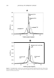
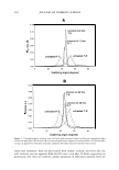
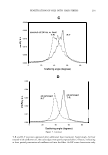
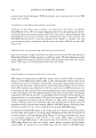
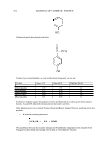
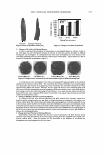
CATIONIC CONDITIONING COMPOUNDS 325 conditioner-treated hair fibers were cross-sectioned each time with a clean stainless steel blade and mounted in small holders with the cross sections facing the spectrometer at a slight tilt. ION MASS SPECTRA Ion mass spectra were collected to isolate characteristic positive/negative ions of the cationics. Positive and negative static TOF SIMS spectra were acquired from several locations on each of the cationic conditioning compounds and from the surface of the hair-fiber cross section. The sampling depth of TOF SIMS is only -1 monolayer for molecular fragment ions and one to three monolayers for atomic species. Since the sampling depth of TOF SIMS is only approximately one molecular layer, only the low-molecular-weight, highly mobile, components in the surface are detected. The higher-molecular-weight compounds are more difficult, if not impossible, to ionize with the 69 Ga + liquid metal ion gun. Therefore, one has to look at the low-molecular weight fragments of the high-molecular-weight compounds. Detecting the fragments, in turn, is indicative of the presence of high-molecular-weight compounds. Positive and negative mass spectra are plotted as the number of secondary ions detected (y-axis, counts) versus the mass-to-charge ratio of the ions (x-axis, m/z). IMAGING Imaging/mapping the presence of cationics within the fiber cross section. Once the characteristic or unique positive or negative ions (atomic species or low-molecular-weight fractions) of the conditioning compounds were established, their diffusion into the fiber cross section was mapped. RES UL TS AND DISCUSSION CHARACTERISTIC POSITIVE AND NEGATIVE IONS OF CET AB Characteristic positive ions of CETAB are C 3 H 8 N + at 58 m/z, (CH 2 \NH 2 + at 114, 128, 142, 156, 170, 184, and 198 m/z, and C 19 H4 2 N + at 284.33 m/z. Although mass C 3 H 8 N+ at 58 m/z is relatively intense, it is probably not a good ion for imaging, since it can be formed from a wide variety of amines and may, therefore, not necessarily be indicative of CETAB if other amines exist. The positive ion C1 9 H4 2 N + at 284.33 m/z is the best ion and will be used for imaging, since it is intense and is likely to be free of mass interference. In combination with this latter ion, imaging of C 3 H 8 N + at 58 m/z will support the presence of CETAB as well. 79 Br- and 81 Br - are characteristic negative ions of CET AB. Because the organic portion of CETAB forms strong positive ions, it tends to form weak negative ions. However, since the presence of sulfates also produces a peak at 81 m/z due to HSO 3 - , that peak is not ideal for mapping CETAB. Therefore, the negative 79 Br- ion seems to be the best for imaging CETAB through the negative ions.
Purchased for the exclusive use of nofirst nolast (unknown)
From: SCC Media Library & Resource Center (library.scconline.org)