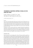
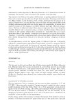
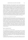
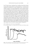
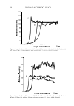
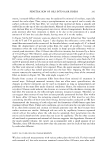
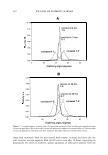
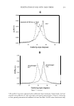
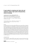
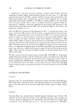
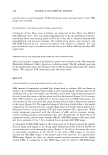
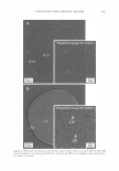
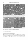
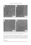
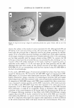
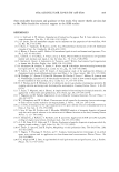
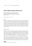
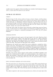
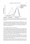
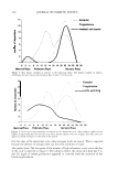
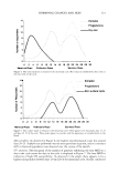
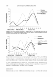
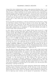
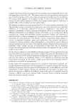
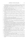
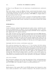
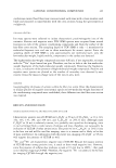
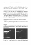
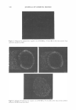
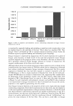
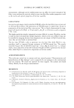
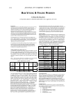
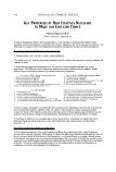
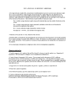
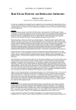
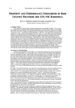
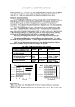
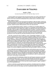
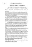
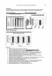
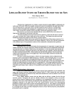
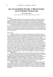
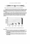
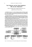
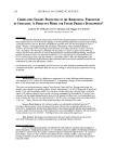
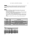
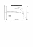
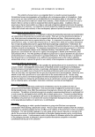
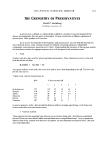
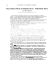
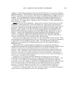
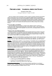
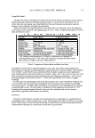
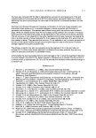
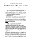
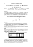
2005 ANNUAL SCIENTIFIC SEMINAR Dye base Dye base with HCE Figure 1 Photo of dyed hair tresses (20') 1. Change in The Color of Coloring Mixtures • l .5 • s::. 11.50 11.00 10.50- 10.00 9.50 1.00 8.50 8.00 7.50 7.00 Smln 10mln 20mln Dyeing Time (minutes) Figure 2 Change In red Index of dyed hair In order to understand the mechanism of enhancement in coloring performance by addition of HCE in the color base, the color development process of mixtures of color base and the developer at different mixing time was investigated. It is obseryed that the initial color development rate in the mixture of the color base containing HCE was slower than that of the mixture without HCE. T yp ical photos at 2 and I 0 minutes of mixing time are presented in Figure 3. Control Base (2') HCE Base (2') Control Base (10') HCE Base (10') Figure 3 Change In color of mixtures of color bases and developer at different mixing time It is clear that the color development in the mixture containing HCE appeared lighter in the emulsion compared to the emulsion without HCE. This could mean that the dye intermediates and coupling agents were protected in the emulsion at the beginning, which would allow the individual components to remain separated until diffuse into the hair. Therefore, the hair dyed with the hair color containing HCE could allow more of the dye intermediate and coupling agents to diffuse into the hair cortex, react inside the hair, thus locking in more color rather than laying on top of the hair. As a result, this may explain why the hair dyed with the hair color containing HCE showed a much better color uptake, fast coloring rate, and final richer color inside the hair cortex. J. Change in Em11lsion Str11ct11re of Coloring Mixt11res It is observed that the color mixture (emulsion) with HCE showed more uniformly dispersed oil droplets, smaller average droplet size, and faster oil droplet-breaking rate compared to the regular color mixture without HCE. Since most of dye intermediates and coupling agent are oil-soluble and prefer to stay in the oil phase, smaller number of oil droplets, more dye intermediates and couplers are distributed outside oil droplets and less chance to interact each other to form complexes outside the hair after oxidation. This means that when the color mixture (emulsion) was applied on hair, dye intermediates and coupling agent in the HCE color mixture had more and better chance to contact with and diffuse into hair, and therefore to enhance the coloring perf onnance. 4. Change in pH and Viscosity of Coloring Mixtures Experimental resuhs also indicated that the color mixture (emulsion) with HCE showed slower increases in pH value and viscosity in the initial stage of mixing (less IO') compared to the regular color mixture without HCE. These two factors are also favourable in the diffusion of un-reacted dye intermediates and coupling agents into the hair cortex. 375

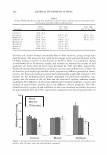
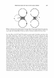
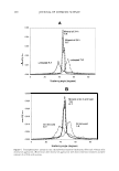
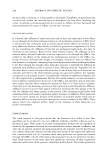
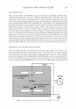
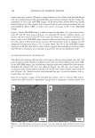
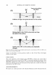
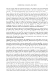
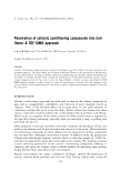
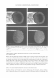
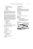
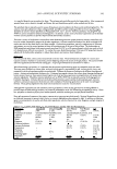
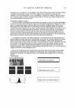
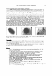
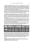
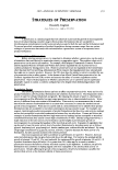
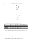
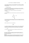
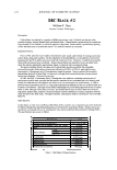
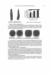
376 JOURNAL OF COSMETIC SCIENCE IN VITRO DERMAL ABSORPTION AND METABOLISM OF D&C RED No. 17 Camille T. Haynes, Ph.D., Robert L. Bronaugh, Ph.D. and Jeffrey J. Yourick, Ph.D. Office of Cosmetics and Colors, Food & Drug Administration, Laurel, MD 20708 Introduction D&C Red No. 17 is approved for use in externally applied drug and cosmetic applications, in amounts consistent with good manufacturing practice. Possible cosmetic uses of D&C Red No. 17 include skin and hair care preparations and suntan products ( 1 ). Concerns about the safety of this color additive ( 1-[ 4- phenylazophenylazo ]-2-naphthol (PAN) is the primary color constituent) have been raised due to potential metabolic cleavage of PAN to yield 4-aminoazobenzene. It was therefore of interest to examine the skin penetration of PAN and to determine if this compound is metabolized in viable skin. Figure 1: Structure of PAN, the primary color constituent of D&C Red No. 17 Methodology In vitro skin absorption studies were conducted in flow-through diffusion cells as previous described (2). Studies were conducted with either freshly obtained viable pig skin or human cadaver skin. The skin was dosed with a commercially available sunscreen product that contained D&C Red No. 17 ( 15 µg/rnl) that was spiked with a"tracer amount of 14 C-PAN. PAN metabolism in viable porcine skin was investigated using HPLC methods. Samples of homogenized viable porcine skin were extracted with ethyl acetate (1:2 vol:vol), which was separated from the aqueous layer and concentrated under a N2 stream. Results Only a small amount of 14 C-PAN was absorbed through human and porcine skin in 24 h (Table 1). Only 0.05% of the applied dose was found in the receptor fluid with human skin. The time course of absorption shows similar absorption between human and pig skin with a significant increase in pig skin absorption in the 24 h sample. Most of the PAN penetrating into the skin remained in the skin. Total penetration was similar in human (10.5 %) and porcine (13.2 %) skin. Table 1: PAN penetration in human and porcine skin after 24 hours (% total applied dose) Human Porcine Receptor fluid 0.05±0.0l 0.53±0.15 Skin content 10.5±1.7 12.6±1.2 Total applied 10.5±1.7 13.2±2.1 dose penetrated Wash 91.8±11.7 91.6±4.7 Recovery 102.4±11.1 104.2±4.9 Values are mean± SEM for human (n=7) and porcine (n=16) skin Because of the large amount of PAN found in the skin at 24 h, extended studies were conducted for 72 h (with a skin wash at 24 h) to determine if additional PAN would be absorbed (Table 2). Receptor fluid levels increased slightly with human skin and were now similar to pig skin values. Porcine skin levels of PAN were unchanged when 24 and 72 h values were compared. Human skin levels also did not decrease during the extended study. It appears that the levels of PAN penetrating into the skin are unable to readily diffuse out of the skin into the receptor fluid even in the extended absorption study.
Purchased for the exclusive use of nofirst nolast (unknown)
From: SCC Media Library & Resource Center (library.scconline.org)