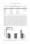
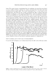
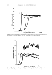
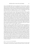
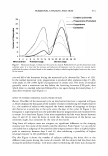
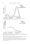
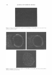
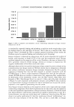
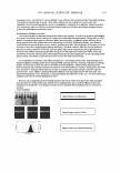
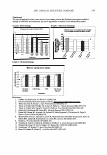
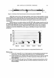
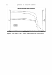
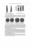
HORMONAL CHANGES AND SKIN 319 lining of the uterus (endometrium) is shed, causing menstrual bleeding. Days 7 to 14 are the follicular or proliferative phase, and days 15 to 28 are the luteal or secretory phase when the egg is released (ovulation). In the normal menstrual cycle, progesterone is produced during ovulation. Generally, during days 1-6 of the cycle there is less than 100 ng/dl of progesterone in the blood. During days 7-14 the level rises to 20-150 ng/dl, and on days 15-28 there is a peak of 250-2,800 ng/dl of progesterone in the blood (22). A similar trend was observed in this study, as observed in Figure 1. It is clear that the subjects used in these studies were within the normal range of monthly hormonal fluctuation. During early follicular development, circulating estradiol levels are relatively low. About one week before ovulation, levels begin to increase, at first slowly, then rapidly. The levels generally reach a maximum one day before the luteinizing hormone (LH) peak. After this peak and before ovulation, there is a marked and precipitous fall. During the luteal phase, estradiol rises to a maximum 5-7 days after ovulation and returns to baseline shortly before menstruation (22). In these studies, the skin barrier was the weakest between days 22 and 26. A weak barrier is defined as having fewer layers and/or weaker cohesivity of the layers of the stratum corneum. Skin thickness and echodensity has been reported to change during the spontaneous menstrual cycle (4). Studies conducted by Eisenbeiss et al. (4) report a statistically significant increase in the skin thickness from phase A (2-4 days) to phase B (12-14 days), but not from phase B to phase C (21-23 days). In studies conducted by Harvell et al. (5 ), TEWL was higher on the day of minimal estrogen/progesterone secretion as compared to the day of maximal estrogen secretion on both back (p = 0.03 7) and forearm (p = 0.021) sites, suggesting that the skin barrier function is less complete on the days just prior to the onset of the menses as compared to the days just prior to ovulation. Significant differences in baseline blood flow also existed for the day of maximal estrogen secretion as compared to the day of maximal progesterone secretion, with higher baseline blood flow recorded on the day of maximal progesterone secretion on both the back (p = 0.021) and forearm (p = 0.009) sites (2). Since all subjects in this study were not "stingers," there was only a slight trend toward elevated neuronal response between days 2 and 12 of the cycle. Neuronal responses, like pain symptoms of many disorders, are reported to vary with menstrual stage. Studies conducted by Giamberardino et al. (7) indicate that menstrual phase dysmenorrhea status can have interacting effects on pain thresholds. Skin response to a challenge with sodium lauryl sulfate has been found to be significantly stronger at day 1 than at days 9 through 11 in the menstrual cycle (8). The influence of the menstrual cycle on skin-prick test reactions to histamine, morphine, and allergen indicate a significant increase in weal-and-flare size to histamine, morphine, and parietaria on days 12 to 16 of the cycle, corresponding to ovulation and peak estrogen levels (24). In this study, sting response appeared to correspond more to skin dryness, since skin was also driest between day 1 and day 6 of the cycle. Estrogens have an important function in many components of human skin, including the epidermis, dermis, vasculature, hair follicle, and the sebaceous, eccrine, and apocrine glands (25). Estrogens have significant roles in skin aging, pigmentation, hair growth, sebum production, and skin cancer (25). Estrogen improves the physical properties of skin by improving water retention and the quality of vascularization. In addition,

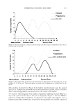
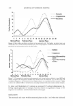
320 JOURNAL OF COSMETIC SCIENCE estrogens have been reported to improve the extracellular matrix responsible for the tone and appearance of the skin (26). The lowest production of progresterone and estrogen was on the first few days of the cycle, when skin appeared to be the most dry. In addition, skin surface lipids appeared to be highest on days 16-20 of the hormonal cycle. The pattern of skin oiliness appears to follow the estrogen release pattern, indicating a possible effect of this hormone on skin surface lipids. The highest microbial count was around days 16 to 22 of the monthly hormonal cycle. The microflora resident on human skin show great inter-individual and intra-individual differences. It is essentially composed of micrococci, staphylococci, and aerobic and anaerobic coryneforms, as well as pityrosporum species, which, in accordance with the different environment in the different regions of the body, are in a steady state. With increasing age, human skin microflora undergo qualitative changes: the streptococci, which are found in infants, disappear and coryneform bacteria occur, which are mainly responsible for odor production (27). Human "native" intracellular sebum, before se cretion, is composed of squalene, waxes, and triglycerides. Once secreted, the sebum is colonized by various xenobiots whose development is controlled by several defensive humoral mechanisms and by contact with ambient oxygen. Oxygen and microorganisms transform "native" sebum, the lysis of triglycerides to fatty acids (28). A correlation between bacterial population and sebum production has been implied (29) and is clearly visible in this study. Several subjects exhibited a lower MED and thereby a higher UV susceptibility between days 20 and 28 of the menstrual cycle. Studies conducted by Jemec and Heidenheim (23) indicate an increased UV-induced inflammation following topical application of estro gen, but they observe no significant change in UV response in correlation with the blood levels of estrogen. In this study, a weak barrier follows the maximum systemic production of estradiol and progesterone. In this study, UV susceptibility appears to be concurrent with barrier condition. Al though epidermal stratification and the formation of a stratum corneum has been hy pothesized to provide protection against UV-B radiation (30-31) the differences in UV-B transmission in both stratum corneum and epidermis are too small to account com pletely for variation in MED at the different times of the cycle. It is possible that this variation in MED is due to the combined effect of several factors, including hormonal levels and stratum corneum integrity. REFERENCES (1) E. Sbano, V. Altamura, F. Galasso, and C. Capilungo, Sexual hormones and acne in adolescent women, G. Ital. Dermatol. Venereal., 125, 363-367 (1990). (2) D. M. Lawrence, M. Katz, T. W. Robinson, M. C. Newman, H. H. McGarrigle, M. Shaw, and G. C. Lacheli'n, Reduced sex hormone binding globulin and derived free testosterone levels in women with severe acne, Clin. Endocrinol., 15, 87-91 (1981). (3) R. Linse and J. Hadlich, Dermatitis as an expression of progesterone hypersensitivity, Zentralbl. Gynakol., 100, 926-930 (1978). (4) C. Eisenbeiss, J. Welzel, and W. Schmeller, The influence of female sex hormones on skin thickness: Evaluation using 20 MHz sonography, Br.]. Dermatol., 139, 462-467 (1998). (5) J. Harvell, I. Hussona-Saeed, and H. I. Maibach, Changes in transepidermal water loss and cutaneous blood flow during the menstrual cycle, Contact Dermatitis, 27, 294-301 (1992). (6) L. Bungum, K. Kvernebo, P. Oian, and J.M. Maltau, Laser doppler-recorded reactive hyperaemia in the forearm skin during the menstrual cycle, Br.]. Obstet. Gynaecol., 103, 70-75 (1996).
Purchased for the exclusive use of nofirst nolast (unknown)
From: SCC Media Library & Resource Center (library.scconline.org)