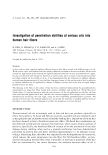
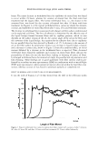
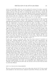
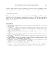
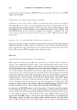
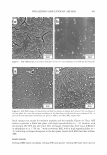
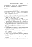
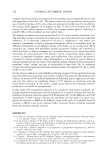
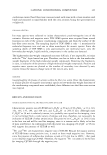
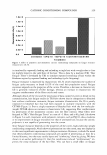
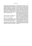
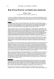
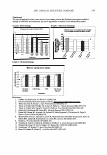
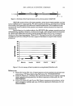
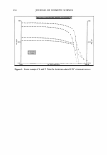
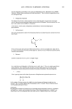
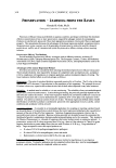
306 JOURNAL OF COSMETIC SCIENCE Figure 9. Optical microscopic images of transversely polished hair planes without ASE (a) and with ASE (b). observe the surfaces or the insides of various materials (10-19). Although both PE and SE belong to the same dry etching system, their principles differ. The PE technique is based upon the principle that a chemically reactive gas plasma generated by radiofre quency or by a microwave electrical discharge can readily react with surface molecules on the target, resulting in the production of volatile molecules (10,13-18), and oxygen gas has been generally used for oxygen plasma etching (OPE). In contrast, the SE technique is based upon the principle that gas ions generated by glow discharge in a low vacuum collide with surface molecules on the target, resulting in sputtering atoms or molecules at the surface (11,12,19), and argon gas has been generally used for ASE. Although there have been studies on the internal structure of hair fibers observed by OPE-SEM (14, 15 ), no corresponding study has been reported using SE (ASE)-SEM to the best of our knowledge. In this study, ASE-SEM images of transversely polished hair planes have been charac terized for the first time. We found that the ASE-SEM images are distinct from OPE SEM images (14, 15) in their three-dimensional structures. Thus, in ASE-SEM, there are highly characteristic images consisting of circular-shaped structures (CP) and slender thread-shaped ones (SP) with convexity in the cortex of transversely polished hair planes. A comparison of ASE-SEM images with TEM images revealed that these convex struc tures correspond to melanin granules and the CMC, respectively, while melanin granules and the CMC in the OPE-SEM images have been reported to be convex and concave, respectively (14,15). Based upon the morphological differences in the CMC between those techniques, it would be of considerable interest to determine what components contribute to the convex formation of SP during ASE. Our observation that SP but not CP disappear following treatment of hair fibers with CHC1/CH 3 OH/water strongly suggests that convex SP are mainly comprised of hair lipids of the CMC since treatment with CHC1/CH 3 OH/water is a typical solvent for removing lipids (20). The suggestion that SP is comprised of hair lipids also supports other experiments in which incubation of the solvent-treated hair fibers with lipids restores the convex formation. Thus, incu-

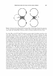
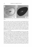
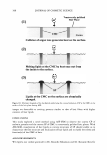
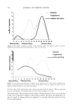
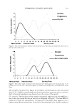
VISUALIZING HAIR LIPIDS BY ASE-SEM 307 bation with fatty acids or wax esters elicits the reappearance of SP, although triolein does not have that effect, which may result from its inability to penetrate into the hair fiber due to its large molecular size. In the mechanism involved in the convex formation of CP, there is a simple etching mechanism as reported previously in several cases (10-19): convex CP are formed due to the great difference between the slower etching of melanin granules and the faster etching of the surrounding proteins. This mechanism is also corroborated by our ob servations that no treatments (such as incubation with solvent, chemical fixation, and thin sectioning) affected the appearance of CP in the hair plane by ASE. As for the mechanism involved in the convex formation of SP, it is of considerable interest to characterize how hair lipids can generate convex structures during ASE. Our findings that the convex SP disappeared after chemical fixation with osmium tetroxide and in 90-nm-thick ultra-thin sections cannot be explained in terms of a simple etching mechanism. Our observation using chemical fixation of hair fibers revealed that fixation with osmium tetroxide, but not with glutaraldehyde, markedly diminished the convex structures of SP. Since osmium tetroxide fixes both proteins and lipids while glutaral dehyde fixes only proteins, it is likely that the existence of mobile lipids is essential for the convex formation of SP during ASE. Another observation using thin sectioning of hair fibers demonstrated that there is no convex formation of SP in ASE-SEM images using 90-nm-thick ultra-thin sections. This result suggests that hair structures with a minimal thickness are essentially required for the convex formation of SP during ASE. In addition, our results using optical microscopy revealed that ASE elicits a remarkable change from a transparent colorless plane to a black opaque one, implying that heat is generated in the hair plane during ASE. We also showed that SP formation is not affected by post-incubation with ethanol, whereas it is completely abolished following pre-incubation with ethanol, which indicates an alteration of the chemical status of hair lipids during ASE. Based upon the above findings, the mechanism(s) involved in the convex formation of SP at the CMC during ASE might be as depicted in Figure 10: (a) joule heat is generated on the surface by violent collisions of argon ions, (b) melting hair lipids at the CMC ooze out from the inside of the hair plane to the surface, and (c) lipids that have oozed out from the CMC are chemically changed by the additional collision energy of argon ions, leading to the final convex formation of SP. The chemical change in hair lipids might be associated with complex reactions such as the polymerization of lipids or the covalent binding of lipids with proteins during ASE. Although the convex SP do not seem to be distinct etching structures, it is plausible that the convexity of SP in ASE-SEM images can be used as a good indicator to visualize the fine structure and localization of hair lipids. In OPE-SEM observations for internal structures of a hair fiber (14, 15), Swift (14) stated that the "cell membrane is etched at far greater rates due to the lowest sulfur content" according to the principle of OPE, which results in the concave structures of the CMC in the surrounding proteins. Al though the location of the CMC in a hair fiber is detectable by OPE-SEM, it is not sufficient to directly visualize the fine structure of hair lipids. This is because the concave structure of the CMC is responsible for the lower content of sulfur within the proteins, but is not relevant to the hair lipids. On the other hand, although conventional TEM also allows the observation of microstructures, including the �-layers and 8-layer of the CMC in a hair fiber, the structures of the �-layers are not directly responsible for the hair lipids. In our experience, even if the hair lipids are lost, the �-layers of the CMC in TEM
Purchased for the exclusive use of nofirst nolast (unknown)
From: SCC Media Library & Resource Center (library.scconline.org)