JOURNAL OF COSMETIC SCIENCE 308 (a) Visual clinical score for assessment of the degree of cellulite (both visual appearance and appear- ance at pinching on the basis of a specifi c photographic reference scale): 0–no observable cellulite 1–no panniculus, some mild depressions 2–some panniculus separated from mild de- pressions 3–several panniculus, separated from medium depressions and 4–broad pan- niculus separated from deep depressions (0 and 4 were considered exclusion criteria). (b) Firmness of the inner thigh (0–very low 1–low 2–moderate 3–good 4–very good). (c) Skin smoothness (0–very low 1–low 2–moderate 3–good 4–very good). (d) Degree of pain at pinching (0–absent 1–mild 2–moderate 3–severe 4–very severe). INSTRUMENTAL EVALUATION The instrumental evaluations were carried out through: (a) Contact thermography for the assessment of the thermographic stage of cellulite (13). At each control, contact thermography was conducted by means of liquid crystal thermographic plates that can detect a temperature range form 28° to 34°C through color visualization (from the coldest to the warmest: black–brown–yellow–green–light blue–pink–dark green–blue). The clinical meaning of contact thermography refl ects the amount of heat that is transmitted to the plate by the contact with the skin. The skin temperature is a clear indication of cutaneous microcirculation, and its variations allow assessing the effi - cacy of vasokinetic treatments: skin temperature increases in the case of vasodilation, in- creased number of open capillaries, and increased local metabolism, while it decreases following vasoconstriction, a decreased number of blood vessels, and a fat tissue increase. One thermographic aspect typical of panniculopathy is dysthermia, with wide hypother- mic areas. The panniculopathy evaluation was based upon the following classifi cation: 0–homogeneous “warm” aspect of the thermographic image 1–dysthermiae 2–“venous” lakes 3–wide cold areas and 4–“cold” aspect of the thermographic image. (b) Morphometric measurements of thigh circumferences (upper, median, and lower third). All cir- cumferences were measured in standardized conditions at the upper, median, and lower third levels of the thigh thanks to a specifi c electro-optical system able to defi ne the vol- unteer’s position. Measurements were taken three times for each site both single values and the median value were recorded. The electro-optical system (composed of a support, a horizontal bar with two laser beams, and a graduated panel in front of the support) al- lowed us to establish precisely the volunteer’s position with respect to the graduated panel behind him (1 mm approximation). In order to determine the coordinates, the subject stood in front of the graduated panel and the operator drew the feet position in order to put him back in the same position during the following visits. Then the laser beam was set to tangentially touch the volunteer’s leg and was pointed on the graduated panel. The same point was then marked on the volunteer’s skin (by means of a dermo- graphic pencil) and the measurement of the thigh circumference was taken. (c) Skin plastoelasticity measured on the inner thigh for the evaluation of elasticizing/fi rming effi - cacy (14). The evaluation of skin plastoelasticity was carried out at the inner thigh level by the means of a dermal torque meter (Diastron LTD). The instrument applies the tech- nique of in vivo torsion by a probe composed of two concentric circles 3 mm apart. The inner circle, spinning slowly, determines a constant torsion on the skin. When the skin resistance reaches 9mNm, the torsion stops. The torsion time is 1 second. The equipment

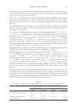
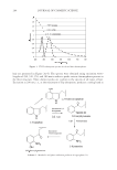
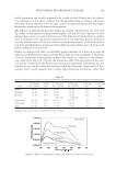
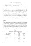
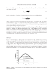
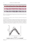
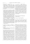
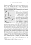
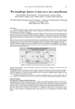
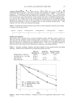
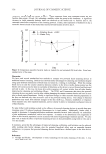
EVALUATION OF ANTI-CELLULITE EFFICACY 309 measures the resulting torsion angle at the maximum of mechanical stimulation and at the end of the stimulus (return phase). For each of the curves, the cutaneous rotation can be measured allowing the defi nition of the following parameters: U • e : immediate extensibility (measured at 0.02 sec) U • f : maximal extensibility (measured at 0.9 sec) U • v : viscoelasticity U • r : immediate elastic return (at 0.02 sec on the return phase) A typical torsion curve example is provided in Figure 1. Among all the available methods to assess cutaneous elasticity, the method involving torsion appears to be one of the most interesting, as it is very sensitive to the variations of the mechanical properties of the stratum corneum (14) (d) Ultrasonography performed on the outer thigh to measure the thickness of the panniculus adiposus (in mm) (15,16). Ultrasonography allowed the measurement of the panniculus adiposus of the upper third of the outer thigh by means of the equipment Body Metrix BX2000 (Genex). In this system, a probe generates high-frequency sound waves, transmitting them within the human body. The waves cross tissues and are refl ected at the tissue inter- faces. By recording the echoes of the refl ected waves, the equipment defi nes the thickness of a certain tissue, and this is made possible by measuring the time it takes for the signal to reach an interface and by multiplying it by the speed of the waves in that specifi c tissue (in the adipose tissue, the speed is around 1400m/s). A watery gel is applied on the probe in order to minimize wave dispersion. (e) Spectrophotometric analysis for the assessment of the activity on surface microcirculation (17). The area of the inner knee represents the most sensitive zone subject to microcirculation variations, where the presence of cellulite is best shown in terms of circulatory stasis. For this reason, the effi cacy of the test product was evaluated with a spectrophotometric mea- surement of the skin color on the inner knee. Spectrophotometric evaluations employed a spectrophotometer for the spectra of visible, infrared, and ultraviolet (λ 300-900 nm) using a tungsten halogen lamp and a deuterium lamp compliant to CIE (Commission Internationale de I’Eclairage). The light source was turned on 30 minutes prior to the use of the equipment in order to stabilize the lamp emissions. The inclination of the probe Figure 1. Skin angular deformation versus time upon application of constant torque. Ue: immediate defor- mation Ur: immediate recovery upon torque switching off.
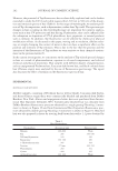
Purchased for the exclusive use of nofirst nolast (unknown)
From: SCC Media Library & Resource Center (library.scconline.org)